
Page 133 - Small Animal Internal Medicine, 6th Edition
P. 133
CHAPTER 5 Congenital Cardiac Disease 105
generated across the stenotic region, as downstream pressure
is normal. The magnitude of this gradient is related to the
VetBooks.ir severity of the obstruction and strength of ventricular
contraction.
Concentric myocardial hypertrophy typically develops in
response to a systolic pressure overload; some dilation of the
affected ventricle also can occur. Ventricular hypertrophy
can impede diastolic filling (by increasing ventricular stiff-
ness) or lead to secondary AV valve regurgitation. Heart
failure results when ventricular diastolic and atrial pressures
are elevated. Cardiac arrhythmias can contribute to the onset
of CHF. Furthermore, the combination of outflow obstruc-
tion, paroxysmal arrhythmias, and/or inappropriate brady-
cardia reflexively triggered by ventricular baroreceptor
stimulation can result in signs of low cardiac output. These
signs are more often associated with severe outflow tract
FIG 5.4 obstruction and include exercise intolerance, syncope, and
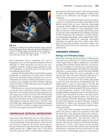
Continuous turbulent flow into the pulmonary artery from the sudden death.
area of the patent ductus arteriosus (arrow) is illustrated in a
color flow Doppler image from the left cranial parasternal SUBAORTIC STENOSIS
position, in an adult female Boston Terrier. Ao, Aorta; PA,
main pulmonary artery; RV, right ventricle. Etiology and Pathophysiology
Subvalvular narrowing caused by a fibrous or fibromuscular
device embolization. Reverse remodeling of LV and LA ring is the most common type of LV outflow stenosis in dogs.
enlargement occurs in most dogs after successful occlusion. Certain larger breeds of dog are predisposed to this defect,
Although LV systolic dimension and function may never including Newfoundlands, Golden Retrievers, and Rottwei-
completely normalize, residual changes generally are clini- lers. SAS is thought to be inherited as an autosomal domi-
cally insignificant. A normal life span can be expected after nant trait with modifying genes that influence its phenotypic
uncomplicated ductal closure. expression; a causative genetic mutation has been identified
Animals with left-sided CHF are treated with furosemide, in Newfoundland dogs. SAS occurs occasionally in cats;
pimobendan, an angiotensin-converting enzyme inhibitor supravalvular lesions have been reported in this species as
(ACEI), rest, and dietary sodium restriction (see Chapter 3). well. Valvular aortic stenosis is reported in Bull Terriers.
Arrhythmias are treated as needed. Ductal closure is recom- The spectrum of SAS severity varies widely; three grades
mended as soon as feasible once CHF is stabilized. Tapering of SAS have been described in Newfoundland dogs. The
or discontinuation of CHF medications may be possible fol- mildest (grade I) causes no clinical signs or murmur and
lowing successful closure. only subtle subaortic fibrous tissue ridging seen on postmor-
If the ductus is not corrected, prognosis depends on ductal tem examination. Moderate (grade II) SAS causes mild clini-
size and the level of pulmonary vascular resistance. Left- cal and hemodynamic evidence of the disease, with an
sided CHF is the eventual outcome for most patients that do incomplete fibrous ring below the aortic valve found at post-
not undergo ductal closure; more than 50% of affected dogs mortem. Dogs with grade III SAS have severe disease and a
die within the first year of life. In animals with pulmonary complete fibrous ring around the outflow tract. Some cases
hypertension and shunt reversal, ductal closure generally is have an elongated, tunnel-like obstruction. Malformation of
contraindicated because the ductus acts as a “pop-off” valve the mitral valve apparatus can exist as well, and a component
for the high right-sided pressures. Ductal ligation in animals of dynamic LV outflow tract obstruction (with or without
with reversed PDA is unlikely to produce improvement and systolic anterior motion of the mitral valve) might be impor-
can lead to acute right ventricular (RV) failure. tant in some dogs.
Unlike many other congenital heart defects, the lesion
itself is not present at birth; rather, patients are born with
VENTRICULAR OUTFLOW OBSTRUCTION abnormal tissue in the subvalvular region of the conotrun-
cal septum that retains the ability to proliferate and undergo
Ventricular outflow obstruction can occur at the semilunar chondrogenic differentiation. The obstructive lesion of
valve, just below the valve (subvalvular), or above the valve SAS therefore develops postnatally during the first several
in the proximal great vessel (supravalvular). SAS and PS are months of life and may continue to worsen until the dog is
most common in dogs and cats. Stenotic lesions impose a fully grown (1-2 years of age). Murmur intensity therefore
pressure overload on the affected ventricle, requiring higher often increases over time and usually increases dynamically
systolic pressure and a slightly longer time to eject blood with exercise or excitement. Because of such factors, as well
across the narrowed outlet. A systolic pressure gradient is as the presence of physiologic murmurs in some animals,