
Page 137 - Small Animal Internal Medicine, 6th Edition
P. 137
CHAPTER 5 Congenital Cardiac Disease 109
VetBooks.ir
A B
C
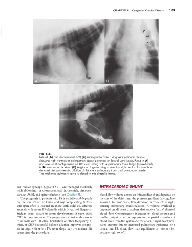
FIG 5.6
Lateral (A) and dorsoventral (DV) (B) radiographs from a dog with pulmonic stenosis,
showing right ventricular enlargement (apex elevation on lateral view [arrowhead in A]
and reverse D configuration on DV view) along with a pulmonary trunk bulge (arrowheads
in B) seen on a DV view. (C) Angiocardiogram using a selective right ventricular injection
demonstrates poststenotic dilation of the main pulmonary trunk and pulmonary arteries.
The thickened pulmonic valve is closed in this diastolic frame.
can reduce syncope. Signs of CHF are managed medically INTRACARDIAC SHUNT
with abdomino- or thoracocentesis, furosemide, pimoben-
dan, an ACEI, and spironolactone (see Chapter 3). Blood flow volume across an intracardiac shunt depends on
The prognosis in patients with PS is variable and depends the size of the defect and the pressure gradient driving flow
on the severity of the lesion and any complicating factors. across it. In most cases, flow direction is from left to right,
Life span often is normal in those with mild PS, whereas causing pulmonary overcirculation. A volume overload is
animals with severe PS often die within 3 years of diagnosis. imposed on all heart chambers that receive “extra” shunted
Sudden death occurs in some; development of right-sided blood flow. Compensatory increases in blood volume and
CHF is more common. The prognosis is considerably worse cardiac output occur in response to the partial diversion of
in animals with TR, atrial fibrillation or other tachyarrhyth- blood away from the systemic circulation. If right heart pres-
mias, or CHF. Successful balloon dilation improves progno- sures increase due to increased pulmonary resistance or a
sis in dogs with severe PS; some dogs may live normal life concurrent PS, shunt flow may equilibrate or reverse (i.e.,
spans after the procedure. become right to left).