
Page 134 - Small Animal Internal Medicine, 6th Edition
P. 134
106 PART I Cardiovascular System Disorders
definitive diagnosis and genetic counseling to breeders can because of the orientation of the aortic arch. The murmur
be difficult. typically is heard over the carotid arteries as well and, in
VetBooks.ir pressure overload and resulting concentric hypertrophy. severe cases, sometimes even radiates to the calvarium.
The severity of the stenosis determines the degree of LV
Aortic regurgitation can produce a diastolic murmur at the
Coronary perfusion is easily compromised in animals with
severe LV hypertrophy. Myocardial capillary density can left base or may be inaudible. Other common physical exam-
ination findings in dogs with moderate to severe stenosis
become inadequate as hypertrophy progresses. Furthermore, include weak and late-rising femoral pulses (pulsus parvus et
the high systolic wall tension, along with coronary narrow- tardus), although concurrent severe aortic regurgitation can
ing, can cause systolic flow reversal in small coronary arter- increase the arterial pulse strength. There may be evidence
ies. These factors contribute to intermittent myocardial of pulmonary edema or arrhythmias.
ischemia and secondary fibrosis. Clinical sequelae include In mild cases, a soft, poorly radiating ejection murmur at
arrhythmias, syncope, and sudden death. Many animals with the left and sometimes right heart base may be the only
SAS also have aortic or mitral valve regurgitation because of abnormality found on physical examination. Low-grade
related malformations or secondary changes; this imposes an functional LV outflow murmurs that are not associated with
additional volume overload on the LV. Left-sided CHF devel- SAS are common in normal Greyhounds, other sighthounds,
ops in some cases. Animals with SAS are thought to be at and Boxers; presence of such physiologic flow murmurs can
higher risk for aortic valve endocarditis because of jet lesion complicate diagnosis of SAS.
injury to the underside of the valve (see p. 132 and Figs. 6.5
and 6.6). Diagnosis
Radiographic abnormalities (see Table 5.2) can be subtle,
Clinical Features especially in animals with mild SAS. The LV can appear
Most patients with SAS are asymptomatic on initial presen- normal or enlarged; mild to moderate LA enlargement is
tation. Clinical signs of fatigue, exercise intolerance or exer- more likely with severe SAS or concurrent MR. Poststenotic
tional weakness, syncope, or sudden death occur in about dilation in the ascending aorta can cause a prominent cranial
one third of dogs with SAS. Low-output signs can result from waist in the cardiac silhouette (especially on a lateral view)
severe outflow obstruction, tachyarrhythmias, or sudden and cranial mediastinal widening. The ECG often is normal,
reflex bradycardia and hypotension resulting from the acti- although evidence of LV hypertrophy (left axis deviation) or
vation of ventricular mechanoreceptors. Signs of left-sided enlargement (tall R waves) can be present. Depression of the
CHF can develop, usually in conjunction with concurrent ST segment in leads II and aVF can occur from myocardial
mitral or aortic regurgitation, other cardiac malformations, ischemia or secondary to hypertrophy; exercise induces
or acquired endocarditis. Dyspnea is the most commonly further ischemic ST-segment changes in some animals. Ven-
reported sign in cats with SAS. tricular tachyarrhythmias are common.
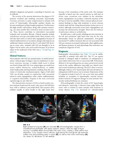
The characteristic murmur of SAS is a harsh systolic ejec- Echocardiography reveals the extent of LV hypertrophy
tion murmur heard near the aortic valve area at the left heart and subaortic narrowing. A discrete tissue ridge below the
base, with or without a precordial thrill. This murmur often aortic valve is evident in many animals with moderate to
radiates equally or more loudly to the right heart base severe disease (Fig. 5.5). Increased LV subendocardial
A B
FIG 5.5
Right parasternal long-axis (A) and subcostal (B) echocardiographic images from a
3-month-old male Rottweiler with severe subaortic stenosis. Note the ridge of tissue
causing a tunnel-like defect (arrow) below the aortic valve, creating a fixed outflow tract
obstruction. Color Doppler reveals turbulent, high-velocity flow through the left ventricular
outflow tract and ascending aorta in systole (B), as well as mild aortic insufficiency (A).
Ao, Aorta; LA, left atrium; LV, left ventricle.