
Page 141 - Small Animal Internal Medicine, 6th Edition
P. 141
CHAPTER 5 Congenital Cardiac Disease 113
VetBooks.ir
A B
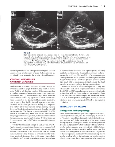
FIG 5.9
Right parasternal long-axis echo images from a 1-year-old male Labrador Retriever with
tricuspid valve dysplasia in diastole (A) and systole (B). The valve annulus appears to be
ventrally displaced; the leaflet tips are tethered to a malformed, wide papillary muscle
(arrows in A). Wide leaflet tip separation in systole (B) caused severe tricuspid
regurgitation and clinical congestive heart failure. LA, Left atrium; LV, left ventricle; RA,
right atrium; RV, right ventricle.
the tricuspid valve under cardiopulmonary bypass has been to hyperviscosity associated with erythrocytosis, including
described in a small number of dogs. Balloon dilation has metabolic and hemostatic abnormalities, seizures, and cere-
occasionally been successful for treating tricuspid stenosis. brovascular accidents. The possibility of a venous embolus
crossing the shunt to the systemic circulation poses another
CARDIAC ANOMALIES danger in these cases. Despite the pressure overload on the
CAUSING CYANOSIS right heart, CHF is rare in cyanotic heart disease; the shunt
provides an alternate pathway for high pressure flow.
Malformations that allow deoxygenated blood to reach the Anomalies that most often cause cyanosis in dogs and
systemic circulation (right-to-left shunts) result in hypox- cats include T of F, PS in conjunction with an intracardiac
emia. Right-to-left shunting requires (1) the presence of an shunt (VSD or ASD), or pulmonary arterial hypertension in
anomalous connection between the systemic and pulmonary conjunction with an intracardiac or extracardiac shunt
circulations, and (2) suprasystemic right heart pressures, (PDA, VSD, or ASD). Other complex but uncommon anom-
usually due to pulmonary hypertension or PS. Visible cya- alies such as transposition of the great vessels or truncus
nosis occurs when the desaturated hemoglobin concentra- arteriosus also send deoxygenated blood to the systemic
tion is greater than 5 g/dL. Arterial hypoxemia stimulates circulation.
increased red blood cell production, leading to a compensa-
tory erythrocytosis that increases oxygen carrying capacity. TETRALOGY OF FALLOT
However, blood viscosity and resistance to flow also rise with
the increase in PCV. Severe erythrocytosis (PCV ≥ 65%) Etiology and Pathophysiology
results in hyperviscosity, which can lead to microvascular T of F is classically defined by its four components: VSD, PS,
sludging, poor tissue oxygenation, intravascular thrombosis, a dextropositioned aorta, and RV hypertrophy. However, T
hemorrhage, and cardiac arrhythmias. Erythrocytosis can of F is actually caused by a single embryologic defect: incom-
become extreme, with a PCV of greater than 80% in some plete rotation and faulty partitioning of the conotruncus
animals. during septation of the great vessels. The malalignment of
Usually the earliest clinical sign in animals with cyanotic the aorta and pulmonary artery with respect to the interven-
heart disease is exercise-induced weakness or syncope. Such tricular septum causes a large nonrestrictive VSD, obstruc-
“hypercyanotic” events occur because exercise stimulates tion of the RV outflow tract (PS), and an aortic root that
systemic vasodilation to increase blood flow to skeletal extends over the right side of the interventricular septum; all
muscles; the resulting decrease in systemic vascular resis- of these components facilitate RV-to-aortic shunting. The PS
tance transiently increases right-to-left shunt volume. Later usually is subvalvular or infundibular, but can involve the
complications of cyanotic heart disease generally are related valve; in some cases, the pulmonary artery is hypoplastic or