
Page 143 - Small Animal Internal Medicine, 6th Edition
P. 143
CHAPTER 5 Congenital Cardiac Disease 115
to those resulting from T of F. The major difference is that the descending aorta, the caudal body receives desaturated
impediment to pulmonary flow occurs at the level of the pul- blood (Fig. 5.10). Rear limb weakness is common in animals
VetBooks.ir monary arterioles rather than at the pulmonic valve. Clinical with reversed PDA.
A murmur typical of the underlying defect(s) might be
manifestations include hypoxemia, cyanosis (worsened with
exercise), RV hypertrophy and enlargement, and erythrocy-
because right and left heart pressures are nearly equivalent,
tosis and its sequelae. Right-sided CHF is uncommon but heard. However, in many cases, no murmur is audible
can develop in response to secondary myocardial failure or minimizing pressure gradient and thus velocity of shunting
tricuspid insufficiency. The right-to-left shunt potentially blood flow. Additionally, high blood viscosity caused by
allows venous emboli to cross into the systemic arterial erythrocytosis minimizes turbulence. There is no continuous
system and cause stroke or other arterial embolization. murmur in patients with reversed PDA. Pulmonary hyper-
tension often causes a loud and “snapping” or split S 2 sound.
Clinical Features Other physical examination findings can include a pro-
The history and clinical presentation of animals with pulmo- nounced right precordial impulse and jugular pulsations.
nary hypertension and shunt reversal are similar to those
associated with T of F. Exercise intolerance, shortness of Diagnosis
breath, syncope (especially in association with exercise or Thoracic radiographs typically reveal right heart enlarge-
excitement), and sudden death are common. Cyanosis might ment, a prominent pulmonary trunk, and tortuous, proxi-
be evident only during exercise or excitement. Intracardiac mally widened pulmonary arteries. A bulge in the descending
shunts cause equally intense cyanosis throughout the body, aorta is common in dogs with reversed PDA. In animals with
whereas a reversed PDA causes cyanosis of the caudal a reversed PDA or VSD, the left heart may be enlarged as
mucous membranes alone (differential cyanosis). In reversed well. The ECG usually suggests RV and sometimes RA
PDA, normally oxygenated blood flows to the cranial body enlargement, with a right axis deviation.
via the brachycephalic trunk and left subclavian artery Echocardiography reveals the RV hypertrophy, intracar-
(from the aortic arch); because the ductus is located in the diac anatomic defects, and sometimes a large ductus, as well
A B
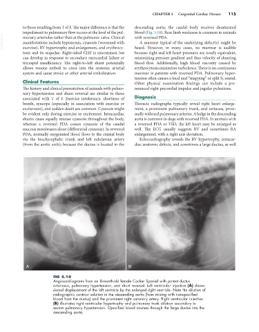
FIG 5.10
Angiocardiograms from an 8-month-old female Cocker Spaniel with patent ductus
arteriosus, pulmonary hypertension, and shunt reversal. Left ventricular injection (A) shows
dorsal displacement of the left ventricle by the enlarged right ventricle. Note the dilution of
radiographic contrast solution in the descending aorta (from mixing with nonopacified
blood from the ductus) and the prominent right coronary artery. Right ventricular injection
(B) illustrates right ventricular hypertrophy and pulmonary trunk dilation secondary to
severe pulmonary hypertension. Opacified blood courses through the large ductus into the
descending aorta.