
Page 203 - Small Animal Internal Medicine, 6th Edition
P. 203
CHAPTER 9 Pericardial Disease and Cardiac Tumors 175
VetBooks.ir
A B
C
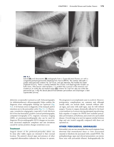
FIG 9.1
Lateral (A) and dorsoventral (B) radiographs from a 5-year-old male Persian cat with a
congenital peritoneopericardial diaphragmatic hernia (PPDH). The cardiac silhouette is
greatly enlarged and contains fat, soft tissue, and gas densities; the trachea has been
displaced dorsally. There is overlap between the cardiac and diaphragmatic borders on
both views. After barium administration, it is evident that a portion of the stomach and
duodenum lie within the pericardial space (C); omental fat and liver also are within the
pericardial sac. In C, the dorsal pleural fold between pericardium and diaphragm is best
appreciated (arrow).
deformity occasionally is present as well. Echocardiography The prognosis in uncomplicated cases is excellent. However,
(or abdominothoracic ultrasonography) helps confirm the perioperative complications are common and, although
diagnosis when radiographic findings are equivocal (Fig. usually mild, can include death. Animals without clini-
9.2). A GI barium series is diagnostic if the stomach and/or cal signs, and some with mild signs, may do well without
intestines are in the pericardial cavity (Fig. 9.1, C). Fluoros- surgery. Trauma to organs chronically adhered to the heart
copy, nonselective angiography (especially if only falciform or pericardium is of concern during attempted repositioning.
fat or liver has herniated), positive contrast peritoneography, Rare sequelae of surgery for PPDH have included pericar-
computed tomography (CT), magnetic resonance imaging dial cyst formation, arrhythmias, and constrictive pericardial
(MRI), or pneumopericardiography also can be used for disease. Overall, long-term survival appears similar between
diagnosis. Electrocardiographic (ECG) changes are inconsis- dogs and cats treated surgically compared with those not
tent; decreased amplitude complexes and axis deviations operated on.
caused by cardiac position changes sometimes occur.
OTHER PERICARDIAL ANOMALIES
Treatment Pericardial cysts are rare anomalies that could originate from
Surgical closure of the peritoneal-pericardial defect can abnormal fetal mesenchymal tissue or from incarcerated
be done after viable organs are returned to their normal omental or falciform fat associated with a small PPDH. The
location. The patient’s clinical signs and presence of other pathophysiologic signs and clinical presentation can mimic
congenital abnormalities influence the decision to operate. those seen with pericardial effusion. Radiographically, the