
Page 207 - Small Animal Internal Medicine, 6th Edition
P. 207
CHAPTER 9 Pericardial Disease and Cardiac Tumors 179
VetBooks.ir
A B
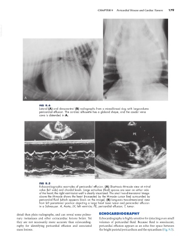
FIG 9.4
Lateral (A) and dorsoventral (B) radiographs from a mixed-breed dog with large-volume
pericardial effusion. The cardiac silhouette has a globoid shape, and the caudal vena
cava is distended in A.
A B
FIG 9.5
Echocardiographic examples of pericardial effusion. (A) Short-axis M-mode view at mitral
valve (left side) and chordal levels. Large echo-free (fluid) spaces are seen on either side
of the heart; the right ventricular wall is clearly visualized. The small two-dimensional image
above the M-mode shows the heart (transected by the M-mode cursor line) surrounded by
pericardial fluid (which appears black on the image). (B) Long-axis two-dimensional view
from left parasternal position depicting a large heart base tumor and pericardial effusion
in a Schnauzer. A, Aorta; LV, left ventricle; PE, pericardial effusion; T, tumor.
detail than plain radiographs, and can reveal some pulmo- ECHOCARDIOGRAPHY
nary metastases and other extracardiac lesions better. Yet Echocardiography is highly sensitive for detecting even small
they are not necessarily more accurate than echocardiog- volumes of pericardial fluid. Because fluid is sonolucent,
raphy for identifying pericardial effusion and associated pericardial effusion appears as an echo-free space between
mass lesions. the bright parietal pericardium and the epicardium (Fig. 9.5).