
Page 209 - Small Animal Internal Medicine, 6th Edition
P. 209
CHAPTER 9 Pericardial Disease and Cardiac Tumors 181
Furthermore, evidence of collapsed lung lobes or pleural neoplastic cells usually are identified easily. Many neoplastic
folds often can be seen within pleural effusion. (and other noninflammatory) effusions have a pH of 7.0 or
VetBooks.ir ELECTROCARDIOGRAPHY greater, whereas inflammatory effusions generally have lower
pH. However, there is too much overlap for pericardial effu-
Although there are no pathognomonic ECG findings, the
culture is done if cytology and pH suggest an infectious or
following abnormalities suggest pericardial effusion but are sion pH to be a reliable discriminator. Pericardial fluid
not seen consistently: small amplitude QRS complexes inflammatory cause. In some patients, fungal titers (for
(<1 mV in dogs), electrical alternans, and ST segment eleva- example, for coccidioidomycosis) or other serologic tests are
tion (epicardial injury current). Electrical alternans is a helpful. Elevated cTnI, either in serum of pericardial fluid,
recurring alteration in the size of the QRS complex (or some- suggests cardiac HSA or other cause of myocardial injury.
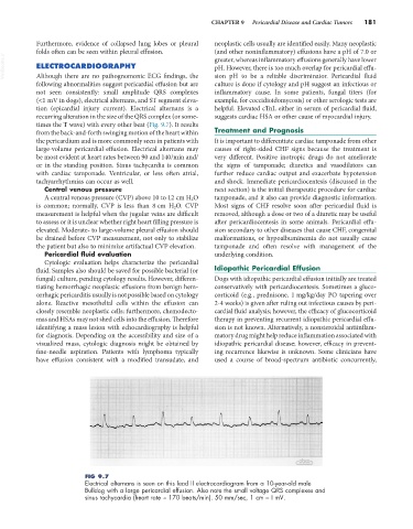
times the T wave) with every other beat (Fig. 9.7). It results
from the back-and-forth swinging motion of the heart within Treatment and Prognosis
the pericardium and is more commonly seen in patients with It is important to differentiate cardiac tamponade from other
large-volume pericardial effusion. Electrical alternans may causes of right-sided CHF signs because the treatment is
be most evident at heart rates between 90 and 140/min and/ very different. Positive inotropic drugs do not ameliorate
or in the standing position. Sinus tachycardia is common the signs of tamponade; diuretics and vasodilators can
with cardiac tamponade. Ventricular, or less often atrial, further reduce cardiac output and exacerbate hypotension
tachyarrhythmias can occur as well. and shock. Immediate pericardiocentesis (discussed in the
Central venous pressure next section) is the initial therapeutic procedure for cardiac
A central venous pressure (CVP) above 10 to 12 cm H 2 O tamponade, and it also can provide diagnostic information.
is common; normally, CVP is less than 8 cm H 2 O. CVP Most signs of CHF resolve soon after pericardial fluid is
measurement is helpful when the jugular veins are difficult removed, although a dose or two of a diuretic may be useful
to assess or it is unclear whether right heart filling pressure is after pericardiocentesis in some animals. Pericardial effu-
elevated. Moderate- to large-volume pleural effusion should sion secondary to other diseases that cause CHF, congenital
be drained before CVP measurement, not only to stabilize malformations, or hypoalbuminemia do not usually cause
the patient but also to minimize artifactual CVP elevation. tamponade and often resolve with management of the
Pericardial fluid evaluation underlying condition.
Cytologic evaluation helps characterize the pericardial
fluid. Samples also should be saved for possible bacterial (or Idiopathic Pericardial Effusion
fungal) culture, pending cytology results. However, differen- Dogs with idiopathic pericardial effusion initially are treated
tiating hemorrhagic neoplastic effusions from benign hem- conservatively with pericardiocentesis. Sometimes a gluco-
orrhagic pericarditis usually is not possible based on cytology corticoid (e.g., prednisone, 1 mg/kg/day PO tapering over
alone. Reactive mesothelial cells within the effusion can 2-4 weeks) is given after ruling out infectious causes by peri-
closely resemble neoplastic cells; furthermore, chemodecto- cardial fluid analysis; however, the efficacy of glucocorticoid
mas and HSAs may not shed cells into the effusion. Therefore therapy in preventing recurrent idiopathic pericardial effu-
identifying a mass lesion with echocardiography is helpful sion is not known. Alternatively, a nonsteroidal antiinflam-
for diagnosis. Depending on the accessibility and size of a matory drug might help reduce inflammation associated with
visualized mass, cytologic diagnosis might be obtained by idiopathic pericardial disease; however, efficacy in prevent-
fine-needle aspiration. Patients with lymphoma typically ing recurrence likewise is unknown. Some clinicians have
have effusion consistent with a modified transudate, and used a course of broad-spectrum antibiotic concurrently,
FIG 9.7
Electrical alternans is seen on this lead II electrocardiogram from a 10-year-old male
Bulldog with a large pericardial effusion. Also note the small voltage QRS complexes and
sinus tachycardia (heart rate ≈ 170 beats/min). 50 mm/sec, 1 cm = I mV.