
Page 204 - Small Animal Internal Medicine, 6th Edition
P. 204
176 PART I Cardiovascular System Disorders
VetBooks.ir
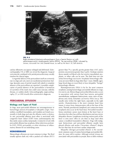
FIG 9.2
Right parasternal short-axis echocardiogram from a female Persian cat with
peritoneopericardial diaphragmatic hernia (PPDH). The pericardium (PERI), indicated by
arrows, surrounds liver and omental tissue, as well as the heart. LV, Left ventricle.
cardiac silhouette can appear enlarged and deformed. Echo- greater than 7%, a specific gravity greater than 1.015, and a
cardiography, CT, or MRI can reveal the diagnosis. Surgical protein concentration greater than 3 g/dL. Cytologic analysis
cyst removal, combined with partial pericardiectomy, usually shows mainly red blood cells, but reactive mesothelial, neo-
resolves the clinical signs. plastic, or other cells can be seen. The fluid does not clot
Congenital defects of the pericardium itself are extremely unless hemorrhage was recent. Neoplastic hemorrhagic effu-
rare in dogs and cats; most are incidental postmortem find- sions are more likely in dogs older than 7 years. Middle-aged,
ings. Sporadic cases of partial (usually left-sided) or complete large-breed dogs are most likely to have idiopathic “benign”
absence of the pericardium are reported. A possible compli- hemorrhagic effusion.
cation of partial absence of the pericardium is herniation Hemangiosarcoma (HSA) is by far the most common
of a portion of the heart; this could cause syncope, embolic neoplasm causing hemorrhagic pericardial effusion in dogs;
disease, or sudden death. Echocardiography, angiocardiog- it is rare in cats. Hemorrhagic pericardial effusion also occurs
raphy, CT, or MRI should allow antemortem diagnosis. in association with various heart base tumors, pericardial
mesothelioma, malignant histiocytosis, some cases of lym-
phoma and, rarely, metastatic carcinoma. HSAs (see p. 185)
PERICARDIAL EFFUSION usually arise within the right heart, especially in the right
auricle. Chemodectoma is the most common heart base
Etiology and Types of Fluid tumor; it arises from chemoreceptor cells at the base of the
In dogs, most pericardial effusions are serosanguineous or aorta. Thyroid, parathyroid, lymphoid, and connective tissue
hemorrhagic and are of neoplastic or idiopathic origin. Tran- neoplasms also occur at the heart base. Pericardial mesothe-
sudates, modified transudates, and exudates are found occa- lioma sometimes causes mass lesions at the heart base or
sionally in both dogs and cats; the effusion rarely is chylous. elsewhere but often has a diffuse distribution and may mimic
In cats, pericardial effusion most often is associated with idiopathic disease. Lymphoma involving various parts of the
congestive heart failure (CHF) from cardiomyopathy, but heart is seen more often in cats than in dogs (and often
this rarely causes cardiac tamponade. A minority of cats with causes a modified transudative effusion). Dogs with malig-
pericardial effusion has neoplasia, feline infectious peritoni- nant histiocytosis and pericardial effusion usually have
tis (FIP), PPDH, pericarditis, or other infectious or inflam- pleural effusion and ascites (“tricavitary effusion”) despite
matory disease for an underlying cause. the fact that they do not have cardiac tamponade.
Idiopathic (benign) pericardial effusion is the second-
HEMORRHAGE most common cause of canine hemorrhagic pericardial effu-
Hemorrhagic effusions are most common in dogs. The fluid sion. Its cause still is unclear. Although several viruses are
usually appears dark red, with a packed cell volume (PCV) associated with pericarditis in people, there is little evidence