
Page 208 - Small Animal Internal Medicine, 6th Edition
P. 208
180 PART I Cardiovascular System Disorders
Large-volume pericardial effusions allow the heart to swing intrapericardial pressure. The RV and RA walls are often well
back and forth within the pericardial sac. Echocardiography visualized and may appear hyperechoic because of the sur-
VetBooks.ir also can identify intrapericardial and intracardiac mass rounding fluid. Better visualization of the heart base and
mass lesions generally is obtained before pericardiocentesis
lesions, as well as abnormal cardiac wall motion, chamber
shape, and other cardiac abnormalities. Clinicians with basic
helpful to do the echocardiographic examination just before
training in echocardiography or just the “thoracic focused is performed. Therefore, if the patient is stable enough, it is
assessment with sonography for trauma” (TFAST) ultra- pericardiocentesis. Careful evaluation of all portions of the
sound examination should be able to identify pericardial and RA and right auricle, RV, ascending aorta, and pericardium
pleural effusions. Especially important in patients with col- itself is important to screen for neoplasia. The left cranial
lapse or respiratory distress, a TFAST examination can reveal parasternal transducer views are especially useful. Some
pericardial effusion and tamponade quickly, even before mass lesions are difficult to visualize. Idiopathic pericardial
radiographs are obtained. Nevertheless, after the patient has effusion is diagnosed only after infectious and neoplastic
been stabilized, a more detailed echocardiographic examina- causes have been excluded. Unfortunately, some mass lesions
tion is warranted to identify and define any mass lesions or are not easily visualized, and mesothelioma without a dis-
other cardiac disease. crete mass lesion cannot be reliably distinguished by nonin-
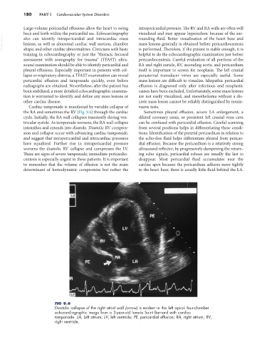
Cardiac tamponade is manifested by variable collapse of vasive tests.
the RA and sometimes the RV (Fig. 9.6) through the cardiac Sometimes pleural effusion, severe LA enlargement, a
cycle. Initially, the RA wall collapses transiently during ven- dilated coronary sinus, or persistent left cranial vena cava
tricular systole. As tamponade worsens, the RA wall collapse can be confused with pericardial effusion. Careful scanning
intensifies and extends into diastole. Diastolic RV compres- from several positions helps in differentiating these condi-
sion and collapse occur with advancing cardiac tamponade, tions. Identification of the parietal pericardium in relation to
and suggest that intrapericardial and intracardiac pressures the echo-free fluid helps differentiate pleural from pericar-
have equalized. Further rise in intrapericardial pressure dial effusion. Because the pericardium is a relatively strong
worsens the diastolic RV collapse and compresses the LV. ultrasound reflector, by progressively dampening the return-
These are signs of severe tamponade; immediate pericardio- ing echo signals, pericardial echoes are usually the last to
centesis is especially urgent in these patients. It is important disappear. Most pericardial fluid accumulates near the
to remember that the volume of effusion is not the main cardiac apex because the pericardium adheres more tightly
determinant of hemodynamic compromise but rather the to the heart base; there is usually little fluid behind the LA.
FIG 9.6
Diastolic collapse of the right atrial wall (arrow) is evident in this left apical four-chamber
echocardiographic image from a 3-year-old female Saint Bernard with cardiac
tamponade. LA, Left atrium; LV, left ventricle; PE, pericardial effusion; RA, right atrium; RV,
right ventricle.