
Page 432 - Small Animal Internal Medicine, 6th Edition
P. 432
404 PART III Digestive System Disorders
The clinician should not assume that constipation, if present, continue to strain after defecating, whereas a constipated
is causing the tenesmus. Severe pain (e.g., that resulting from animal strains before feces are produced. Tenesmus that
VetBooks.ir proctitis) may make the animal refuse to defecate and cause occurs when an animal is in a squatting position often results
from colitis, whereas tenesmus that occurs when an animal is
secondary constipation. Most rectal strictures, perineal
in a semiwalking or partial squatting position usually results
hernias, masses, enlarged prostates, pelvic fractures, and
rectal tumors can be detected during a digital rectal exami- from constipation.
nation. The clinician may need to use two fingers to detect
partial strictures when examining large dogs. Perianal fistu-
lae are usually visible but may be detected only as perirectal CONSTIPATION
thickenings. Next, the clinician expresses the anal sacs and
examines their contents. Finally, the clinician evaluates the Constipation (infrequent and difficult evacuation of feces)
feces to determine whether they are excessively hard or have and obstipation (intractable constipation) have several causes
abnormal contents (e.g., hair, trash). (Box 26.15). The initial use of symptomatic therapy is often
A biopsy should be done of any mass, stricture, or infiltra- successful, but it is important to look for causes because
tive lesion found by rectal examination. A rectal scraping is some problems may become harder to treat if symptomatic
sometimes sufficient (e.g., histoplasmosis), otherwise biopsy therapy masks signs while the underlying disease progresses.
specimens taken with rigid biopsy forceps that include sub- Iatrogenic, dietary, environmental, or behavioral causes
mucosa are preferred. Fine-needle aspiration should be per- should be sought on history. Feces should be examined to
formed on extracolonic masses because abscesses occasionally determine whether they contain plastic, bones, hair, popcorn,
occur. or other such material. Physical and digital rectal examina-
If the clinician is confused by physical examination tions are done to search for rectal obstruction or infiltration.
findings, observing the animal defecate may help define Plain pelvic radiographs can help show whether the animal
the underlying process. Animals with inflammation often has anatomic abnormalities or a previously undetected
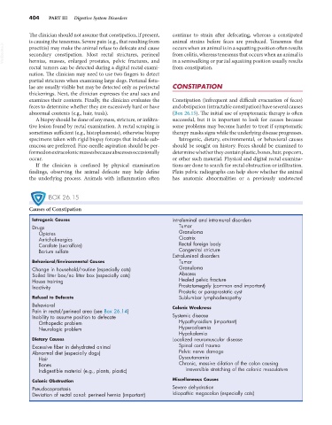
BOX 26.15
Causes of Constipation
Iatrogenic Causes Intraluminal and intramural disorders
Drugs Tumor
Opiates Granuloma
Anticholinergics Cicatrix
Carafate (sucralfate) Rectal foreign body
Barium sulfate Congenital stricture
Extraluminal disorders
Behavioral/Environmental Causes Tumor
Change in household/routine (especially cats) Granuloma
Soiled litter box/no litter box (especially cats) Abscess
House training Healed pelvic fracture
Inactivity Prostatomegaly (common and important)
Prostatic or paraprostatic cyst
Refusal to Defecate Sublumbar lymphadenopathy
Behavioral
Pain in rectal/perineal area (see Box 26.14) Colonic Weakness
Inability to assume position to defecate Systemic disease
Orthopedic problem Hypothyroidism (important)
Neurologic problem Hypercalcemia
Hypokalemia
Dietary Causes Localized neuromuscular disease
Excessive fiber in dehydrated animal Spinal cord trauma
Abnormal diet (especially dogs) Pelvic nerve damage
Hair Dysautonomia
Bones Chronic, massive dilation of the colon causing
Indigestible material (e.g., plants, plastic) irreversible stretching of the colonic musculature
Colonic Obstruction Miscellaneous Causes
Pseudocoprostasis Severe dehydration
Deviation of rectal canal: perineal hernia (important) Idiopathic megacolon (especially cats)