
Page 434 - Small Animal Internal Medicine, 6th Edition
P. 434
406 PART III Digestive System Disorders
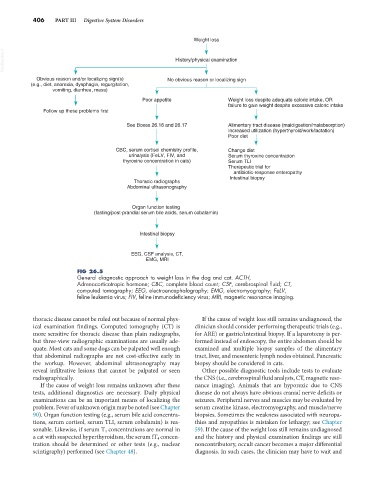
Weight loss
VetBooks.ir History/physical examination
Obvious reason and/or localizing sign(s) No obvious reason or localizing sign
(e.g., diet, anorexia, dysphagia, regurgitation,
vomiting, diarrhea, mass)
Poor appetite Weight loss despite adequate caloric intake, OR
failure to gain weight despite excessive caloric intake
Follow up these problems first
See Boxes 26.16 and 26.17 Alimentary tract disease (maldigestion/malabsorption)
Increased utilization (hyperthyroid/work/lactation)
Poor diet
CBC, serum cortisol chemistry profile, Change diet
urinalysis (FeLV, FIV, and Serum thyroxine concentration
thyroxine concentration in cats) Serum TLI
Therapeutic trial for
antibiotic-response enteropathy
Intestinal biopsy
Thoracic radiographs
Abdominal ultrasonography
Organ function testing
(fasting/post-prandial serum bile acids, serum cobalamin)
Intestinal biopsy
EEG, CSF analysis, CT,
EMG, MRI
FIG 26.5
General diagnostic approach to weight loss in the dog and cat. ACTH,
Adrenocorticotropic hormone; CBC, complete blood count; CSF, cerebrospinal fluid; CT,
computed tomography; EEG, electroencephalography; EMG, electromyography; FeLV,
feline leukemia virus; FIV, feline immunodeficiency virus; MRI, magnetic resonance imaging.
thoracic disease cannot be ruled out because of normal phys- If the cause of weight loss still remains undiagnosed, the
ical examination findings. Computed tomography (CT) is clinician should consider performing therapeutic trials (e.g.,
more sensitive for thoracic disease than plain radiographs, for ARE) or gastric/intestinal biopsy. If a laparotomy is per-
but three-view radiographic examinations are usually ade- formed instead of endoscopy, the entire abdomen should be
quate. Most cats and some dogs can be palpated well enough examined and multiple biopsy samples of the alimentary
that abdominal radiographs are not cost-effective early in tract, liver, and mesenteric lymph nodes obtained. Pancreatic
the workup. However, abdominal ultrasonography may biopsy should be considered in cats.
reveal infiltrative lesions that cannot be palpated or seen Other possible diagnostic tools include tests to evaluate
radiographically. the CNS (i.e., cerebrospinal fluid analysis, CT, magnetic reso-
If the cause of weight loss remains unknown after these nance imaging). Animals that are hyporexic due to CNS
tests, additional diagnostics are necessary. Daily physical disease do not always have obvious cranial nerve deficits or
examinations can be an important means of localizing the seizures. Peripheral nerves and muscles may be evaluated by
problem. Fever of unknown origin may be noted (see Chapter serum creatine kinase, electromyography, and muscle/nerve
90). Organ function testing (e.g., serum bile acid concentra- biopsies. Sometimes the weakness associated with neuropa-
tions, serum cortisol, serum TLI, serum cobalamin) is rea- thies and myopathies is mistaken for lethargy; see Chapter
sonable. Likewise, if serum T 4 concentrations are normal in 59). If the cause of the weight loss still remains undiagnosed
a cat with suspected hyperthyroidism, the serum fT 4 concen- and the history and physical examination findings are still
tration should be determined or other tests (e.g., nuclear noncontributory, occult cancer becomes a major differential
scintigraphy) performed (see Chapter 48). diagnosis. In such cases, the clinician may have to wait and