
Page 433 - Small Animal Internal Medicine, 6th Edition
P. 433
CHAPTER 26 Clinical Manifestations of Gastrointestinal Disorders 405
colonic obstruction (e.g., prostatomegaly, enlarged sublum-
bar lymph node). Ultrasonography is the preferred tech- BOX 26.16
VetBooks.ir nique when looking for infiltrates. A serum biochemistry Causes of Weight Loss
panel may reveal causes of colonic inertia (e.g., hypothyroid-
ism, or rarely hypercalcemia or hypokalemia,).
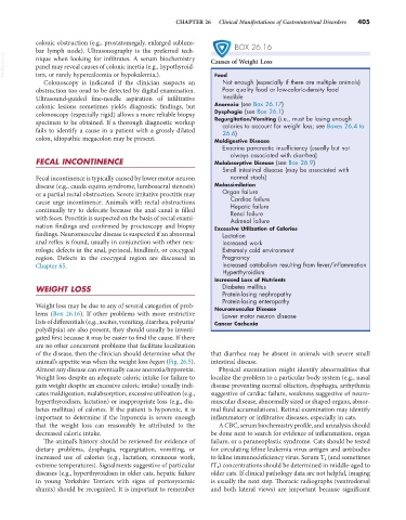
Not enough (especially if there are multiple animals)
Colonoscopy is indicated if the clinician suspects an Food
obstruction too orad to be detected by digital examination. Poor quality food or low-caloric-density food
Ultrasound-guided fine-needle aspiration of infiltrative Inedible
colonic lesions sometimes yields diagnostic findings, but Anorexia (see Box 26.17)
colonoscopy (especially rigid) allows a more reliable biopsy Dysphagia (see Box 26.1)
Regurgitation/Vomiting (i.e., must be losing enough
specimen to be obtained. If a thorough diagnostic workup calories to account for weight loss; see Boxes 26.4 to
fails to identify a cause in a patient with a grossly dilated 26.6)
colon, idiopathic megacolon may be present. Maldigestive Disease
Exocrine pancreatic insufficiency (usually but not
always associated with diarrhea)
FECAL INCONTINENCE Malabsorptive Disease (see Box 26.9)
Small intestinal disease (may be associated with
Fecal incontinence is typically caused by lower motor neuron normal stools)
disease (e.g., cauda equina syndrome, lumbosacral stenosis) Malassimilation
or a partial rectal obstruction. Severe irritative proctitis may Organ failure
cause urge incontinence. Animals with rectal obstructions Cardiac failure
Hepatic failure
continually try to defecate because the anal canal is filled Renal failure
with feces. Proctitis is suspected on the basis of rectal exami- Adrenal failure
nation findings and confirmed by proctoscopy and biopsy Excessive Utilization of Calories
findings. Neuromuscular disease is suspected if an abnormal Lactation
anal reflex is found, usually in conjunction with other neu- Increased work
rologic defects in the anal, perineal, hindlimb, or coccygeal Extremely cold environment
region. Defects in the coccygeal region are discussed in Pregnancy
Chapter 65. Increased catabolism resulting from fever/inflammation
Hyperthyroidism
Increased Loss of Nutrients
WEIGHT LOSS Diabetes mellitus
Protein-losing nephropathy
Protein-losing enteropathy
Weight loss may be due to any of several categories of prob- Neuromuscular Disease
lems (Box 26.16). If other problems with more restrictive Lower motor neuron disease
lists of differentials (e.g., ascites, vomiting, diarrhea, polyuria/ Cancer Cachexia
polydipsia) are also present, they should usually be investi-
gated first because it may be easier to find the cause. If there
are no other concurrent problems that facilitate localization
of the disease, then the clinician should determine what the that diarrhea may be absent in animals with severe small
animal’s appetite was when the weight loss began (Fig. 26.5). intestinal disease.
Almost any disease can eventually cause anorexia/hyporexia. Physical examination might identify abnormalities that
Weight loss despite an adequate caloric intake (or failure to localize the problem to a particular body system (e.g., nasal
gain weight despite an excessive caloric intake) usually indi- disease preventing normal olfaction, dysphagia, arrhythmia
cates maldigestion, malabsorption, excessive utilization (e.g., suggestive of cardiac failure, weakness suggestive of neuro-
hyperthyroidism, lactation) or inappropriate loss (e.g., dia- muscular disease, abnormally sized or shaped organs, abnor-
betes mellitus) of calories. If the patient is hyporexic, it is mal fluid accumulations). Retinal examination may identify
important to determine if the hyporexia is severe enough inflammatory or infiltrative diseases, especially in cats.
that the weight loss can reasonably be attributed to the A CBC, serum biochemistry profile, and urinalysis should
decreased caloric intake. be done next to search for evidence of inflammation, organ
The animal’s history should be reviewed for evidence of failure, or a paraneoplastic syndrome. Cats should be tested
dietary problems, dysphagia, regurgitation, vomiting, or for circulating feline leukemia virus antigen and antibodies
increased use of calories (e.g., lactation, strenuous work, to feline immunodeficiency virus. Serum T 4 (and sometimes
extreme temperatures). Signalments suggestive of particular fT 4 ) concentrations should be determined in middle-aged to
diseases (e.g., hyperthyroidism in older cats, hepatic failure older cats. If clinical pathology data are not helpful, imaging
in young Yorkshire Terriers with signs of portosystemic is usually the next step. Thoracic radiographs (ventrodorsal
shunts) should be recognized. It is important to remember and both lateral views) are important because significant