
Page 456 - Small Animal Internal Medicine, 6th Edition
P. 456
428 PART III Digestive System Disorders
VetBooks.ir
A B
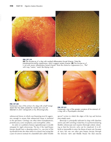
FIG 27.19
(A) The duodenum of a dog with marked inflammatory bowel disease. Note the
pseudomembrane-like appearance, which suggests severe disease. (B) The duodenum of
a cat with severe inflammatory bowel disease. Note the distinctive appearance (i.e., “flat”
with long “cracks,” much like drying mud).
FIG 27.20
Endoscopic view of the antrum of a dog with a ball foreign
object that has been present for months and was not FIG 27.21
detected on plain radiographs or by ultrasonography. Endoscopic view of the greater curvature of the stomach of
a dog with a Physaloptera attached.
submucosal lesion, in which case biopsying must be aggres- spoon”) action in which the edges of the top and bottom
sive enough to ensure that submucosal tissue is included jaws simply meet.
in the specimen. Cytology can often detect histoplasmosis, Ileoscopy is principally indicated in dogs with diarrhea
protothecosis, some neoplasms, and eosinophilic colitis. and in cats with vomiting or diarrhea. It is performed during
An adult or a pediatric human sigmoidoscope is usually flexible colonoscopy and requires thorough colonic cleans-
adequate for rigid colonoscopy. The tip of the rigid biopsy ing so that the ileocolic valve can be visualized. It is dif-
forceps should have a shearing action (i.e., one part of the ficult or impossible to enter the ileum of most cats (because
tip should fit into the other when it is closed, thus acting like of size), but one can often pass biopsy forceps through
a pair of scissors) instead of a clamshell (also called “double the ileocolic valve and blindly biopsy the ileal mucosa