
Page 457 - Small Animal Internal Medicine, 6th Edition
P. 457
CHAPTER 27 Diagnostic Tests for the Alimentary Tract 429
VetBooks.ir
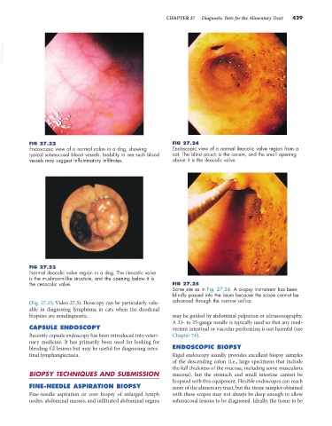
FIG 27.22 FIG 27.24
Endoscopic view of a normal colon in a dog, showing Endoscopic view of a normal ileocolic valve region from a
typical submucosal blood vessels. Inability to see such blood cat. The blind pouch is the cecum, and the small opening
vessels may suggest inflammatory infiltrates. above it is the ileocolic valve.
FIG 27.23
Normal ileocolic valve region in a dog. The ileocolic valve
is the mushroom-like structure, and the opening below it is
the cecocolic valve. FIG 27.25
Same site as in Fig. 27.24. A biopsy instrument has been
blindly passed into the ileum because the scope cannot be
(Fig. 27.25; Video 27.3). Ileoscopy can be particularly valu- advanced through the narrow orifice.
able in diagnosing lymphoma in cats when the duodenal
biopsies are nondiagnostic. may be guided by abdominal palpation or ultrasonography.
A 23- to 25-gauge needle is typically used so that any inad-
CAPSULE ENDOSCOPY vertent intestinal or vascular perforation is not harmful (see
Recently capsule endoscopy has been introduced into veteri- Chapter 74).
nary medicine. It has primarily been used for looking for
bleeding GI lesions but may be useful for diagnosing intes- ENDOSCOPIC BIOPSY
tinal lymphangiectasia. Rigid endoscopy usually provides excellent biopsy samples
of the descending colon (i.e., large specimens that include
the full thickness of the mucosa, including some muscularis
BIOPSY TECHNIQUES AND SUBMISSION mucosa), but the stomach and small intestine cannot be
biopsied with this equipment. Flexible endoscopes can reach
FINE-NEEDLE ASPIRATION BIOPSY more of the alimentary tract, but the tissue samples obtained
Fine-needle aspiration or core biopsy of enlarged lymph with these scopes may not always be deep enough to allow
nodes, abdominal masses, and infiltrated abdominal organs submucosal lesions to be diagnosed. Ideally, the tissue to be