
Page 452 - Small Animal Internal Medicine, 6th Edition
P. 452
424 PART III Digestive System Disorders
with weight loss of uncertain cause and to better define cats Fecal α 1 -protease inhibitor can be measured in feces and
with known small intestinal disease (cobalamin-deficient is a marker for GI protein loss. Clinically this test is rarely
VetBooks.ir cats can experience metabolic complications). If the serum indicated but could be helpful when trying to distinguish
whether hypoalbuminemia is at least partly due to a protein-
cobalamin is low in a patient with weight loss of unknown
cause, small intestinal disease may be responsible. B-complex
or hepatic insufficiency. The test is performed by the GI Lab
vitamin supplementation may cause an increased serum losing enteropathy in a patient with known renal protein loss
cobalamin concentration. at Texas A&M University.
Dietary folate is absorbed in the small intestine. Excessive Tests for Pythium insidiosum are available. ELISA tests for
numbers of bacteria in the upper small intestine sometimes antibodies and PCR testing for antigen can be done at the
synthesize and release folate, causing the serum concentra- College of Veterinary Medicine, Louisiana State University,
tions to be increased. Likewise, severe intestinal mucosal Baton Rouge, LA 70803.
disease may decrease absorption, causing lower serum
concentrations. B-complex vitamin supplementation may
increase serum folate concentrations. Like cobalamin, folate ENDOSCOPY
concentrations are insensitive for intestinal disease and not
specific for any particular intestinal disease. Because bright Endoscopy can be cost effective in animals with chronic
light degrades cobalamin, samples should be frozen and kept vomiting, diarrhea, or weight loss if cases are selected care-
in the dark during storage and transport. fully and the endoscopist is accomplished. It permits rapid
exploration of selected sections of the alimentary tract and
OTHER SPECIAL TESTS FOR mucosal biopsy without the need for a thoracotomy or
ALIMENTARY TRACT DISEASE laparotomy. Although excellent for detecting morphologic
Antibodies to acetylcholine receptors should be measured if changes (e.g., masses, ulcers, obstruction), it is insensitive for
the clinician is looking for localized myasthenia, which is a revealing abnormal function (e.g., esophageal weakness).
potential cause of dysphagia or esophageal weakness (see p. Rigid endoscopy of the colon is easier to perform and less
454). Increased titers to such antibodies are strongly sugges- expensive than flexible endoscopy, and it provides excellent
tive of myasthenia gravis, even if there are no systemic signs. biopsy samples. Flexible endoscopes allow one to examine
False-positive results are rare. Serum can be sent to Dr. the ileocolic and cecocolic valve areas as well as the ascend-
Diane Shelton (Comparative Neuromuscular Laboratory, ing and transverse colons, areas that cannot be inspected
Basic Science Building, University of California at San Diego, with a rigid endoscope. Flexible instruments are expensive
La Jolla, CA 92093-0612) for this analysis. and require time and commitment to become proficient in
Measurement of antibodies to 2M muscle fibers can be their use. One is limited by how far the instrument can be
helpful in dogs with suspected masticatory muscle myositis advanced. Expertise is required to obtain diagnostic tissue
(see p. 451). These antibodies are typically not found in dogs samples without excessive artifacts.
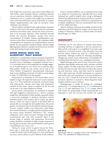
with polymyositis, whereas most dogs with masticatory Esophagoscopy is useful in looking for esophageal
myositis do have them. Serum is required for the test and tumors (Fig. 27.9), foreign objects (Fig. 27.10), inflammation
can be sent to Dr. Diane Shelton for testing. (Fig. 27.11), and obstruction (Fig. 27.12). Foreign objects
Serum gastrin concentrations are measured in animals and cicatrix are preferentially treated endoscopically. It is
with signs suggestive of gastrinoma (i.e., chronic vomiting, important to enter the stomach and retroflex the scope’s
weight loss, and diarrhea in older animals, especially if there
is concurrent esophagitis or duodenal ulceration). Gastrin
stimulates gastric acid secretion and is trophic for gastric
mucosa. Serum for assay of gastrin is harvested from an
animal after an overnight fast and rapidly frozen. The serum
gastrin concentration may be increased in animals with gas-
trinoma, a gastric outflow tract obstruction, renal failure,
short-bowel syndrome, or atrophic gastritis and those receiv-
ing proton pump inhibitors (histamine-2 [H 2 ] receptor
antagonists cause a lesser increase). Resting serum gastrin
concentrations may vary, with occasional values in the
normal range in animals with gastrinoma. Provocative
testing should be considered in dogs strongly suspected of
having gastrinoma but with normal baseline serum gastrin
concentrations (see Chapter 49).
Testing for urease activity in gastric mucosa for Helico-
bacter spp. is rarely done anymore. Helicobacter spp. can FIG 27.9
usually be found easily by histology. For more information Endoscopic view of a polypoid mass in the esophagus of a
on urease activity, please see prior editions of this textbook. Chow. This represents an adenocarcinoma.