
Page 448 - Small Animal Internal Medicine, 6th Edition
P. 448
420 PART III Digestive System Disorders
VetBooks.ir
A B
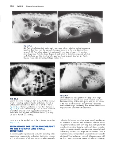
FIG 27.5
(A) Plain lateral abdominal radiograph from a dog with an intestinal obstruction causing
intestinal distention. Note the markedly increased diameter of the small intestinal lumen
(arrows). (B) Plain lateral abdominal radiograph from a dog with peritonitis causing
physiologic ileus. Note the lesser degree of small intestinal distention compared with that
in A. The large gas-filled structure is the gastric pylorus (arrows). (Courtesy Dr. Kenita
Rogers, Texas A&M University, College Station, TX.)
FIG 27.7
Lateral abdominal radiograph from a dog with a large
FIG 27.6 granuloma caused by pythiosis. Small intestinal loops are
Lateral abdominal radiograph from a dog that had an acute displaced dorsally and caudally (small arrows). The border
onset of vomiting, abdominal pain, and shock. There is a of the mass is not discernible except where it displaces
uniform intestinal distention that is not as great as that in small intestinal loops. The finding of a dilated intestinal loop
Fig. 27.5, A. However, distention is more than that seen in (long arrows) is consistent with obstruction.
Fig. 27.5, B. Some intestinal loops have assumed a vertical
orientation (arrows), which suggests the existence of an
obstruction. This dog had a mesenteric volvulus. (Courtesy
Dr. Susan Yanoff, U.S. Military.)
there to be a few gas bubbles in the peritoneal cavity (see evaluating the hepatic parenchyma; and identifying abdomi-
Fig. 32.1, B). nal neoplasia in animals with abdominal effusion. Ultra-
sonography is much more revealing than radiography in
INDICATIONS FOR ULTRASONOGRAPHY animals with minimal body fat that have little or no radio-
OF THE STOMACH AND SMALL graphic contrast in the abdomen. However, very dehydrated
INTESTINES animals may be difficult to image with ultrasound, and it is
Ultrasonography is particularly useful for detecting intus- easy to miss small foreign objects (especially in the stomach/
susceptions, pancreatitis, abdominal infiltrative disease, intestines if food and gas are present). Ultrasonography will
and small amounts of effusion not seen radiographically; not detect bony changes and modest microhepatica that are