
Page 449 - Small Animal Internal Medicine, 6th Edition
P. 449
CHAPTER 27 Diagnostic Tests for the Alimentary Tract 421
detected by radiographs. The skill of the ultrasonographer examination. The clinician should not hesitate to sedate the
determines the usefulness of the technique. dog if it is hyperactive or hyperventilating.
VetBooks.ir Technique Findings
Before ultrasonography is performed, the abdominal hair is
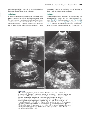
plain radiographs detect, plus gastric and intestinal infil-
usually clipped to improve the quality of the examination. Ultrasonography detects almost any soft tissue change that
This is not necessary in animals with minimal hair. Because trates (Fig. 27.8, A), intussusceptions (see Fig. 27.8, B),
air in the stomach or intestines limits the usefulness of ultra- enlarged lymph nodes (see Fig. 27.8, C), masses (see Fig.
sonography, exercise, drugs (e.g., some narcotics) that cause 27.8, D), some radiolucent foreign objects, and small amounts
hyperventilation, and enemas should be avoided before the of free peritoneal fluid that radiographs cannot detect. If
A B
C D
FIG 27.8
(A) Ultrasonographic image of two sections of small intestine from a cat with an
alimentary tract lymphoma. The normal intestine on the right is 2.8 mm thick (see the two
“+’s” noted as D2), whereas the abnormal intestine on the left is 4.5 mm thick (D1)
because of neoplastic infiltrates. (B) Ultrasonographic image of an ileocolic
intussusception that was not obvious on plain abdominal radiographs. There are two
intestinal walls (small arrows) seen on each side of the lumen (large arrow). (C) An
enlarged mesenteric lymph node in a dog caused by lymphoma, seen by ultrasonography.
The lymph node was not detected on radiographs or by abdominal palpation. (D)
Ultrasound image of the gastric antrum from a dog with benign gastric polyps. One polyp
can be seen (arrows) protruding into the gastric lumen. (Courtesy Dr. Linda Homco,
Cornell University, Ithaca, NY.)