
Page 453 - Small Animal Internal Medicine, 6th Edition
P. 453
CHAPTER 27 Diagnostic Tests for the Alimentary Tract 425
VetBooks.ir
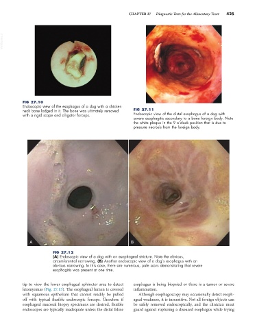
FIG 27.10
Endoscopic view of the esophagus of a dog with a chicken
neck bone lodged in it. The bone was ultimately removed FIG 27.11
with a rigid scope and alligator forceps. Endoscopic view of the distal esophagus of a dog with
severe esophagitis secondary to a bone foreign body. Note
the white plaque in the 9 o’clock position that is due to
pressure necrosis from the foreign body.
A B
FIG 27.12
(A) Endoscopic view of a dog with an esophageal stricture. Note the obvious,
circumferential narrowing. (B) Another endoscopic view of a dog’s esophagus with an
obvious narrowing. In this case, there are numerous, pale scars demonstrating that severe
esophagitis was present at one time.
tip to view the lower esophageal sphincter area to detect esophagus is being biopsied or there is a tumor or severe
leiomyomas (Fig. 27.13). The esophageal lumen is covered inflammation.
with squamous epithelium that cannot readily be pulled Although esophagoscopy may occasionally detect esoph-
off with typical flexible endoscopic forceps. Therefore if ageal weakness, it is insensitive. Not all foreign objects can
esophageal mucosal biopsy specimens are desired, flexible be safely removed endoscopically, and the clinician must
endoscopes are typically inadequate unless the distal feline guard against rupturing a diseased esophagus while trying