
Page 446 - Small Animal Internal Medicine, 6th Edition
P. 446
418 PART III Digestive System Disorders
VetBooks.ir
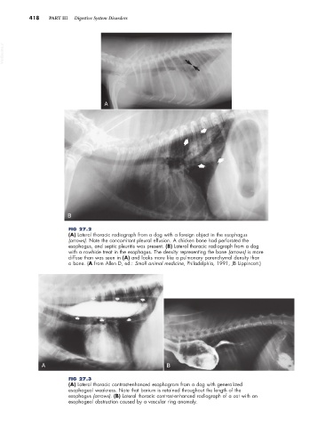
A
B
FIG 27.2
(A) Lateral thoracic radiograph from a dog with a foreign object in the esophagus
(arrows). Note the concomitant pleural effusion. A chicken bone had perforated the
esophagus, and septic pleuritis was present. (B) Lateral thoracic radiograph from a dog
with a rawhide treat in the esophagus. The density representing the bone (arrows) is more
diffuse than was seen in (A) and looks more like a pulmonary parenchymal density than
a bone. (A from Allen D, ed.: Small animal medicine, Philadelphia, 1991, JB Lippincott.)
A B
FIG 27.3
(A) Lateral thoracic contrast-enhanced esophagram from a dog with generalized
esophageal weakness. Note that barium is retained throughout the length of the
esophagus (arrows). (B) Lateral thoracic contrast-enhanced radiograph of a cat with an
esophageal obstruction caused by a vascular ring anomaly.