
Page 447 - Small Animal Internal Medicine, 6th Edition
P. 447
CHAPTER 27 Diagnostic Tests for the Alimentary Tract 419
in which case reexamination of the esophagus with endos-
copy or fluoroscopy or both are required.
VetBooks.ir IMAGING OF THE STOMACH AND
SMALL INTESTINE
INDICATIONS FOR RADIOGRAPHIC
IMAGING OF THE ABDOMEN WITHOUT
CONTRAST MEDIA
Common indications for plain abdominal radiography
include vomiting, acute abdomen, constipation, hyporexia,
abdominal pain, abdominal enlargement, abdominal disten-
tion, or a mass. Plain radiographs are rarely beneficial in
animals with a marked abdominal effusion (the fluid obliter-
ates serosal detail) or with chronic diarrhea. Plain abdominal
radiographs can be especially helpful in detecting radiodense
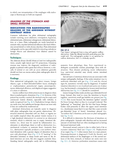
foreign objects and alimentary tract dilation caused by FIG 27.4
Plain lateral radiograph from a dog with gastric outflow
obstruction. obstruction. Note the dilated stomach protruding past the
costal arch. This radiographic pattern is not sensitive for
Techniques outflow obstruction, but it is relatively specific.
The clinician always should obtain at least two radiographic
views, usually right lateral and VD projections. Cleansing
enemas may improve the diagnostic usefulness of radio- anatomic from physiologic ileus. Even experienced ra-
graphs in patients with a great deal of feces; however, a criti- diologists occasionally confuse physiologic ileus with ob-
cally ill animal or one with an acute abdomen generally struction. Thus diseases producing severe inflammation
should not have an enema unless plain radiographs show it (e.g., parvoviral enteritis) may closely mimic intestinal
is necessary. obstruction.
Special types of intestinal obstructions are associated with
Findings unique radiographic findings. If the entire intestinal tract is
Plain abdominal radiographs may detect masses, foreign uniformly distended with gas (Fig. 27.6) and the clinical
objects, a gas- or fluid-distended hollow viscus, misshapen signs fit, mesenteric volvulus may be diagnosed. If marked
or emphysematous parenchymal organs, pneumoperito- intestinal distention is very localized and seems out of place
neum, abdominal effusions, and displaced organs suggestive (e.g., has herniated), a strangulated or incarcerated intestinal
of a mass or adhesion. obstruction (see Fig. 31.11) should be considered.
Gastric outflow tract obstruction is easy to diagnose when Linear foreign bodies rarely produce gas-distended bowel
there is marked gastric distention (Fig. 27.4). However, if the loops. Instead, they tend to cause the intestines to bunch
patient has recently vomited, the stomach may be empty and together; sometimes small gas bubbles are present (see Fig.
contracted. Gastric dilation, especially with volvulus, is 31.12). This occurs because the intestines “gather” around
easily recognized (see Fig. 30.3). Radiodense foreign objects the linear foreign object as they try to propel it aborad. This
are easily seen, but radiolucent foreign objects are seen only “gathering” or “bunching” plus the fact that linear foreign
if they are outlined by swallowed air. bodies tend primarily to affect the upper small intestines
Intestinal obstructions are typically easier to diagnose (i.e., duodenum) mean that it is rare that they cause severely
with plain radiographs than are gastric obstructions. Ob- gas-distended loops of bowel. Sometimes pleated (i.e.,
structed intestines distended with air, fluid, or ingesta are “accordion-like”) intestines can be seen on plain radiographs
not readily emptied when the patient vomits (unless it is (see Fig. 31.12).
a high duodenal obstruction) in contrast to an obstructed It is difficult to determine the thickness of intestines on
stomach, which is relatively easy to empty. Intestinal dis- plain radiographs. Animals with diarrhea and an increased
tention (i.e., ileus) may be caused by inflammation (i.e., amount of intestinal fluid are often misdiagnosed as having
adynamic or physiologic ileus) as well as obstruction (i.e., thickened intestinal walls.
mechanical, occlusive, or anatomic ileus). Anatomic ileus Decreased serosal contrast is due to either lack of fat or
(i.e., obstruction) typically produces a non-uniform intes- excessive abdominal fluid (see Chapter 34). Displacement
tinal distention with a greater degree of distention than is of an organ (Fig. 27.7) often means there is a mass present.
seen with physiologic ileus (Fig. 27.5). If “stacking” of the Pneumoperitoneum is diagnosed if both the thoracic and
distended intestines or sharp bends and turns in the di- abdominal surfaces of the diaphragm or if the serosal sur-
lated intestines are seen, this also suggests anatomic ileus. faces of the liver, stomach, or kidneys are easily seen (see
Standing lateral radiographs rarely aid in differentiating Fig. 32.1, A). However, perforation sometimes causes only