
Page 445 - Small Animal Internal Medicine, 6th Edition
P. 445
CHAPTER 27 Diagnostic Tests for the Alimentary Tract 417
pneumothorax), an isotonic iodine contrast medium should rawhide treats) are relatively radiolucent (Fig. 27.2). An
be used. However, the only purpose of such a study is to esophageal perforation sometimes causes pneumothorax,
VetBooks.ir localize the perforation. If the clinician already knows where pneumomediastinum, or a pleural/mediastinal effusion.
Contrast-enhanced esophagrams should be considered
the leakage is likely to be (e.g., there is a bone foreign body
in animals with unidentified thoracic masses because many
in the esophagus), contrast radiographs are of dubious value.
Perforations may not be obvious if the foreign body that esophageal tumors radiographically resemble pulmonary
caused them is filling and occluding the defect. parenchymal masses (see Fig. 29.8). Contrast-enhanced
esophagrams may also show that structures that seem-
Findings ingly involve the esophagus actually do not. An obstruc-
Esophageal dilation, foreign objects, soft tissue densities, tion is suggested on contrast-enhanced esophagrams if the
spondylosis suggestive of spirocercosis, and hiatal hernia barium column terminates abruptly as it travels caudally;
may often be identified on plain films. An air-filled esopha- weakness usually causes contrast to be retained throughout
gus is not always diagnostic of pathologic esophageal weak- the esophagus or the weakened segment of the esophagus
ness. Although it is tempting to use plain radiograph findings (Fig. 27.3). A partial obstruction is suggested by retention
as the basis for the diagnosis of esophageal disease when of barium-impregnated food but not liquid barium (see
there is an “obvious” abnormality, it is possible to misinter- Fig. 29.7).
pret plain films or miss abnormalities that a barium contrast– A barium contrast study will not always reveal a hiatal
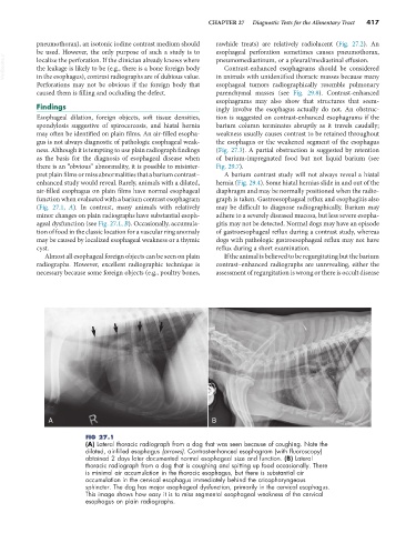
enhanced study would reveal. Rarely, animals with a dilated, hernia (Fig. 29.4). Some hiatal hernias slide in and out of the
air-filled esophagus on plain films have normal esophageal diaphragm and may be normally positioned when the radio-
function when evaluated with a barium contrast esophagram graph is taken. Gastroesophageal reflux and esophagitis also
(Fig. 27.1, A). In contrast, many animals with relatively may be difficult to diagnose radiographically. Barium may
minor changes on plain radiographs have substantial esoph- adhere to a severely diseased mucosa, but less severe esopha-
ageal dysfunction (see Fig. 27.1, B). Occasionally, accumula- gitis may not be detected. Normal dogs may have an episode
tion of food in the classic location for a vascular ring anomaly of gastroesophageal reflux during a contrast study, whereas
may be caused by localized esophageal weakness or a thymic dogs with pathologic gastroesophageal reflux may not have
cyst. reflux during a short examination.
Almost all esophageal foreign objects can be seen on plain If the animal is believed to be regurgitating but the barium
radiographs. However, excellent radiographic technique is contrast–enhanced radiographs are unrevealing, either the
necessary because some foreign objects (e.g., poultry bones, assessment of regurgitation is wrong or there is occult disease
A B
FIG 27.1
(A) Lateral thoracic radiograph from a dog that was seen because of coughing. Note the
dilated, air-filled esophagus (arrows). Contrast-enhanced esophagram (with fluoroscopy)
obtained 2 days later documented normal esophageal size and function. (B) Lateral
thoracic radiograph from a dog that is coughing and spitting up food occasionally. There
is minimal air accumulation in the thoracic esophagus, but there is substantial air
accumulation in the cervical esophagus immediately behind the cricopharyngeous
sphincter. The dog has major esophageal dysfunction, primarily in the cervical esophagus.
This image shows how easy it is to miss segmental esophageal weakness of the cervical
esophagus on plain radiographs.