
Page 602 - Small Animal Internal Medicine, 6th Edition
P. 602
574 PART IV Hepatobiliary and Exocrine Pancreatic Disorders
essential finding. A search should then be conducted for a
possible cause of obstruction by carefully examining the
0
VetBooks.ir 1 small intestine, liver, and pancreas for evidence of inflamma-
tion or neoplasia. Biliary tract rupture can present in a
similar way and should be ruled out by identifying and ana-
lyzing any free abdominal fluid; cats with biliary rupture
2 have a high concentration of bilirubin in the fluid. FNA of
bile from the gallbladder under ultrasonographic guidance
CHD x
3 should be avoided or approached with great care if EBDO is
suspected or confirmed because of the high risk of leakage
4 caused by the increased pressure. In these cats it is preferable
to aspirate bile during surgery. It may be necessary to under-
take an exploratory laparotomy to assess bile duct patency
and the cause of the obstruction. Hemostatic function should
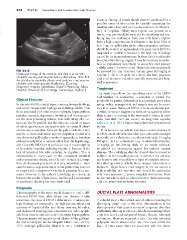
FIG 35.8 be assessed first, and vitamin K therapy given (0.5 mg/kg of
Ultrasound image of the common bile duct in a cat with vitamin K 1 SC or IM q12h for 3 days). The liver, pancreas,
choleliths causing extrahepatic biliary obstruction. Note that and small intestine should be carefully inspected and biop-
the bile duct is markedly dilated and contains a radiodense
cholelith with distal acoustic shadowing. (Courtesy sied as necessary.
Diagnostic Imaging Department, Queen’s Veterinary School
Hospital, University of Cambridge, Cambridge, England.) Treatment
Treatment depends on the underlying cause of the EBDO
and whether the obstruction is complete or partial. The
Clinical Features prognosis for partial obstructions is surprisingly good when
In cats with EBDO, clinical signs, clinicopathologic findings, using medical management, and surgery may not be neces-
and survey radiographic findings are indistinguishable from sary in all cases. Studies of EBDO in acute-on-chronic pan-
those associated with other severe cholestatic hepatopathies; creatitis in humans suggest that medical management rather
jaundice, anorexia, depression, vomiting, and hepatomegaly than surgery or stenting is the treatment of choice in most
are the main presenting features. Cats with biliary obstruc- cases and that there are usually no long-term sequelae
tion can also be painful, and the clinician should be aware (Abdallah et al., 2007). Similar studies have not been reported
of subtle signs because cats tend to hide their pain. If biliary in cats.
obstruction is complete, feces will be pale or acholic. There If the feces are not acholic and there is some evidence of
may be a cranial abdominal mass on palpation because of a bile flow into the duodenum and no pain, cats can be managed
very distended gallbladder or underlying neoplasia, but often medically with a choleretic (ursodeoxycholic acid, 15 mg/kg
abdominal palpation is normal (other than the hepatomeg- PO q24h) and an antioxidant (e.g., S-adenosylmethionine,
aly). Cats with EBDO are at particular risk of malabsorption 20 mg/kg, or 200-400 mg daily on an empty stomach)
of fat-soluble vitamins, including vitamin K, because of the to protect the hepatocytes against bile-induced oxidant
lack of intestinal bile salts reducing fat digestion. This is damage. The underlying disorder should also be treated as
compounded in many cases by the concurrent intestinal outlined in the preceding section. However, if the cat does
and/or pancreatic disease, which further reduces fat absorp- not improve after several days or signs of complete obstruc-
tion. As discussed previously, it is very important in these tion develop, such as acholic feces, surgical intervention is
cases to assess coagulation times before performing biopsies indicated. Major biliary tract surgery in the cat carries a
or surgery and to supplement vitamin K parenterally as nec- high morbidity and mortality, and should be undertaken
essary. However, to the author’s knowledge, no correlation only when necessary to relieve complete obstruction. More
between the results of hemostasis profiles and development minor procedures such as sphincterotomy, cholecystectomy,
of postbiopsy bleeding has been established. and stent placement are better tolerated.
Diagnosis
Ultrasonography is the most useful diagnostic tool to dif- DUCTAL PLATE ABNORMALITIES
ferentiate EBDO from other biliary tract diseases in cats;
sometimes, the cause of EBDO is determined. Clinicopatho- The ductal plate is the distinct layer of cells surrounding the
logic findings are nonspecific; the high concentrations and developing portal triad in the fetus. Abnormalities in the
activities of hepatocellular and biliary enzymes, bilirubin, development of this cause a variety of congenital disorders
and cholesterol resulting from cholestasis are indistinguish- particularly affecting intrahepatic bile ducts including biliary
able from those in cats with other cholestatic hepatopathies. cysts (see later) and congenital hepatic fibrosis. Although
Ultrasonography will usually reveal dilation of the gallblad- uncommon, these are reported in cats. Cats with inherited
der and extrahepatic and intrahepatic biliary trees (see Fig. polycystic kidney disease often also have cysts within the
35.8), although gallbladder dilation is not a consistent or liver. In some cases, these are associated with the classic