
Page 603 - Small Animal Internal Medicine, 6th Edition
P. 603
CHAPTER 35 Hepatobiliary Diseases in the Cat 575
mutation for polycystic kidney disease, and in others, they
are not (Guerra et al., 2015). Congenital hepatic fibrosis has
VetBooks.ir also been reported in cats, and Persian cats appear to be
over-represented (Bosje et al., 1988; Zandvliet et al., 2005).
Affected cats usually present with signs of portal hyperten-
sion including ascites, lethargy and vomiting, and HE. Alter-
natively, lesions may be discovered incidentally on ultrasound.
Portal hypertension is unusual in cats and should raise the
suspicion of ductal plate abnormality particularly in areas
where liver fluke is not endemic. Inexperienced pathologists
may erroneously diagnose some cases of ductal plate abnor-
mality as biliary cirrhosis. Cytokeratin staining of liver sec-
tions helps highlight the classic marked proliferation of small
bile ducts seen in the bridging fibrosis in congenital hepatic
fibrosis.
Affected cats are treated symptomatically for portal
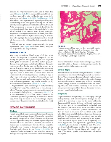
hypertension (see Chapter 36 for more details). Prognosis FIG 35.9
can be good with life-long treatment. Photomicrograph of liver specimen from a cat with hepatic
cystadenoma. Note the multiple cystic spaces lined with
BILIARY CYSTS biliary epithelium (H&E stain). (Courtesy Pathology
Department, Queen’s Veterinary School Hospital, University
Most cystic lesions in the feline liver are of bile duct origin of Cambridge, Cambridge, England.)
and may be congenital or acquired. Congenital cysts are
usually multiple and often present as part of a congenital
ductal plate abnormality as described earlier, polycystic chronic inflammatory process in another organ (e.g., chronic
disease of several organs, including the kidneys. The cystic gingivitis), which is thought to be the driving force for the
contents are clear. Persian cats and Persian crosses are at formation of the inflammatory amyloid.
increased risk. Cysts may be an incidental finding on imaging,
particularly if they are small, but large cysts can cause clinical Clinical Signs
signs as a result of destruction of hepatic tissue and also Affected cats usually present with signs of anemia and hypo-
compression of surrounding bile ducts resulting in signs of tension related to rupture of the hepatic capsule and hemoab-
biliary tract obstruction (see earlier). Treatment is not indi- domen. These cats are predisposed to hepatic rupture because
cated if they are small and nonprogressive, but if they are the liver is enlarged and rigid, and therefore easily damaged
large and causing problems, they may be treated surgically with normal trauma, such as jumping. Affected cats usually
by removal or omentalization (Friend et al., 2001). exhibit lethargy, anorexia, pale mucous membranes, bound-
Acquired hepatic cysts may be single or multiple and may ing pulse, and heart murmur secondary to the anemia but
be small or very large. The contents may be clear, bloody, or rarely any specific signs of liver disease. There may be hepa-
bilious. They may occur secondary to trauma, inflammation, tomegaly on abdominal palpation.
or neoplasia (including biliary cystadenomas; Fig. 35.9) or
in rare cases caused by liver flukes. Therapy depends on the Diagnosis
cause, but surgical management may be necessary if they are Diagnosis relies on histopathology of a liver biopsy; although
large. Cysts may also occur in the form of large, irregular clinicopathologic and ultrasonographic findings are sup-
sacculations of the extrahepatic bile duct (choledochal cysts). portive, it is important to rule out the major differential
diagnoses of FIP, hepatic lipidosis, and hepatic lymphoma.
The transient anemia resolves as blood is reabsorbed from
HEPATIC AMYLOIDOSIS the abdomen (autotransfusion). There are mild to marked
increases in ALT activity and globulin concentration, but
Etiology there are rarely increases in ALP and GGT levels, which
Hepatic amyloidosis is an uncommon but apparently emerg- helps differentiate amyloidosis from biliary tract disease and
ing cause of liver disease in cats. Historically, amyloidosis lipidosis. On ultrasonography, amyloidosis can resemble
was usually recognized as a familial disease in Abyssinian lymphoma and lipidosis, with hepatomegaly and a gener-
cats, which predominantly affects the kidneys although liver alized increase in hepatic parenchymal echogenicity or a
involvement is not uncommon. However, amyloidosis in mixed hypoechoic and hyperechoic appearance (Beatty
Siamese cats is predominantly hepatic. Hepatic amyloido- et al., 2002), but no dilation of the biliary tract. FNA cytol-
sis has also been reported in domestic shorthairs, oriental ogy is not helpful because amyloid does not appear on
shorthairs, and a Devon Rex cat (Beatty et al., 2002). The the aspirate. Therefore hepatic biopsy, after careful evalu-
amyloid in familial and sporadic cases is amyloid A (inflam- ation of hemostasis profiles, is the recommended method
matory), and in sporadic cases there is usually an underlying of diagnosis.