
Page 601 - Small Animal Internal Medicine, 6th Edition
P. 601
CHAPTER 35 Hepatobiliary Diseases in the Cat 573
biliary tract disease, such as dilation of the bile ducts. In one compressive or intraluminal obstructive lesions, but diseases
case fluke infestation also caused acquired polycystic disease often cause EBDO through a combination of these mecha-
VetBooks.ir of the biliary system (Xavier et al., 2007). nisms; for example, cholangitis may result in a combina-
Ova may be found in the feces using the formalin-ether
tion of extraluminal compression by associated edema and
sedimentation method (Box 35.3). However, shedding of
bile. Therefore it is more practically helpful to divide the
eggs is sporadic; also, eggs will not be present if the fluke inflammation and intraluminal obstruction by inspissated
infestation has resulted in a complete biliary obstruction. causes into common and less common causes (Box 35.4).
The most reliable way of demonstrating flukes and eggs is Several studies have shown inflammation of the small
with bile aspirates. intestine, pancreas, biliary tract, or a combination of these
(known as triaditis) to be the most common cause of EBDO
Treatment in cats; neoplasias of the biliary tract or pancreas are the
The ideal and most effective treatment regimen for feline next most common cause. Dysfunction of the sphincter of
liver flukes remains controversial. Currently, the most com- Oddi because of adjacent duodenal inflammation or neo-
monly recommended treatment is praziquantel (20 mg/kg plasia has also been recently reported in cats and may be
SC q24h for 3 days). The prognosis for recovery in severely more common than appreciated because of difficulties in
affected cats is poor. diagnosis (Furneaux, 2010). Choleliths are uncommon in
cats. Those reported in the literature are usually cholesterol
CHOLECYSTITIS or calcium salts, or a mixture of these, and are associated
Cholecystitis refers to inflammation of the gallbladder. Neu- with cholangitis. They are variably radiodense depend-
trophilic cholecystitis is frequently seen in cats but rarely in ing on the amount of calcium in the stone, but they are
dogs. It may occur alone or in combination with neutrophilic easily visualized using ultrasonography (Fig. 35.8). Two of
cholangitis. Ultrasonographically, the gallbladder wall often the three cases of bilirubin choleliths reported in the litera-
appears thickened and sometimes irregular; there may be ture were from Somali cats with pyruvate kinase deficiency,
sludging of the bile and/or choleliths. Clinical signs, diagno- and it was assumed that they were secondary to chronic
sis, and treatment are similar to those of neutrophilic chol- hemolysis (Harvey et al., 2007). Therefore finding bilirubin
angitis (see earlier). Chronic thickening of the gall bladder choleliths in a cat should stimulate a search for underlying
wall may be an indications for surgical cholecystectomy hemolytic disease.
since bacterial infections recur in these patients. Lympho-
cytic cholecystitis is also occasionally recognized and is
treated like lymphocytic cholangitis (see earlier).
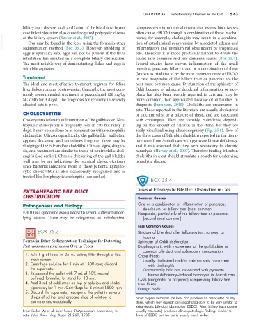
BOX 35.4
EXTRAHEPATIC BILE DUCT Causes of Extrahepatic Bile Duct Obstruction in Cats
OBSTRUCTION Common Causes
Pathogenesis and Etiology One or a combination of inflammation of pancreas,
duodenum, or biliary tree (most common)
EBDO is a syndrome associated with several different under- Neoplasia, particularly of the biliary tree or pancreas
lying causes. These may be categorized as extraluminal (second most common)
Less Common Causes
BOX 35.3 Stricture of bile duct after inflammation, surgery, or
trauma
Formalin-Ether Sedimentation Technique for Detecting Sphincter of Oddi dysfunction
Platynosomum concinnum Ova in Feces Diaphragmatic with involvement of the gallbladder or
common bile duct and subsequent compression
1. Mix 1 g of feces in 25 mL saline; filter through a fine Cholelithiasis
mesh screen. Usually cholesterol and/or calcium salts concurrent
2. Centrifuge solution for 5 min at 1500 rpm; discard with cholangitis
the supernate. Occasionally bilirubin, associated with pyruvate
3. Resuspend the pellet with 7 mL of 10% neutral kinase deficiency–induced hemolysis in Somali cats
buffered formalin; let stand for 10 min. Cysts (congenital or acquired) compressing biliary tree
4. Add 3 mL of cold ether on top of solution and shake Liver flukes
vigorously for 1 min. Centrifuge for 3 min at 1500 rpm. Foreign body
5. Discard the supernate, resuspend the pellet in several
drops of saline, and prepare slide of solution to Note: Sepsis distant to the liver can produce an associated biliary
examine microscopically. stasis, which may appear clinicopathologically to be very similar to
extrahepatic bile duct obstruction (EBDO). Also, biliary tract rupture
From Bielsa LM et al: Liver flukes (Platynosomum concinnum) in (usually traumatic) produces clinicopathologic findings similar to
cats, J Am Anim Hosp Assoc 21:269, 1985. those of EBDO but the cat is usually much sicker.