
Page 596 - Small Animal Internal Medicine, 6th Edition
P. 596
568 PART IV Hepatobiliary and Exocrine Pancreatic Disorders
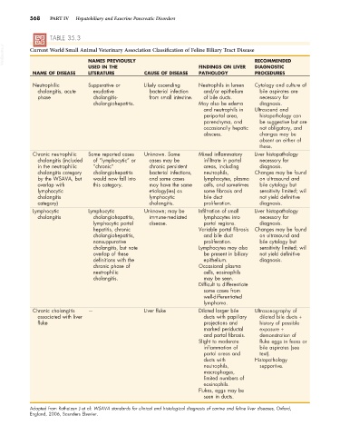
TABLE 35.3
VetBooks.ir Current World Small Animal Veterinary Association Classification of Feline Biliary Tract Disease RECOMMENDED
NAMES PREVIOUSLY
USED IN THE FINDINGS ON LIVER DIAGNOSTIC
NAME OF DISEASE LITERATURE CAUSE OF DISEASE PATHOLOGY PROCEDURES
Neutrophilic Suppurative or Likely ascending Neutrophils in lumen Cytology and culture of
cholangitis, acute exudative bacterial infection and/or epithelium bile aspirates are
phase cholangitis- from small intestine. of bile ducts. necessary for
cholangiohepatitis. May also be edema diagnosis.
and neutrophils in Ultrasound and
periportal area, histopathology can
parenchyma, and be suggestive but are
occasionally hepatic not obligatory, and
abscess. changes may be
absent on either of
these.
Chronic neutrophilic Some reported cases Unknown. Some Mixed inflammatory Liver histopathology
cholangitis (included of “lymphocytic” or cases may be infiltrate in portal necessary for
in the neutrophilic “chronic” chronic persistent areas, including diagnosis.
cholangitis category cholangiohepatitis bacterial infections, neutrophils, Changes may be found
by the WSAVA, but would now fall into and some cases lymphocytes, plasma on ultrasound and
overlap with this category. may have the same cells, and sometimes bile cytology but
lymphocytic etiology(ies) as some fibrosis and sensitivity limited; will
cholangitis lymphocytic bile duct not yield definitive
category) cholangitis. proliferation. diagnosis.
Lymphocytic Lymphocytic Unknown; may be Infiltration of small Liver histopathology
cholangitis cholangiohepatitis, immune-mediated lymphocytes into necessary for
lymphocytic portal disease. portal regions. diagnosis.
hepatitis, chronic Variable portal fibrosis Changes may be found
cholangiohepatitis, and bile duct on ultrasound and
nonsuppurative proliferation. bile cytology but
cholangitis, but note Lymphocytes may also sensitivity limited; will
overlap of these be present in biliary not yield definitive
definitions with the epithelium. diagnosis.
chronic phase of Occasional plasma
neutrophilic cells, eosinophils
cholangitis. may be seen.
Difficult to differentiate
some cases from
well-differentiated
lymphoma.
Chronic cholangitis — Liver fluke Dilated larger bile Ultrasonography of
associated with liver ducts with papillary dilated bile ducts +
fluke projections and history of possible
marked periductal exposure +
and portal fibrosis. demonstration of
Slight to moderate fluke eggs in feces or
inflammation of bile aspirates (see
portal areas and text).
ducts with Histopathology
neutrophils, supportive.
macrophages,
limited numbers of
eosinophils.
Flukes, eggs may be
seen in ducts.
Adapted from Rothuizen J et al: WSAVA standards for clinical and histological diagnosis of canine and feline liver diseases, Oxford,
England, 2006, Saunders Elsevier.