
Page 593 - Small Animal Internal Medicine, 6th Edition
P. 593
CHAPTER 35 Hepatobiliary Diseases in the Cat 565
1996) and a more recent study identified hypoproteinaemia,
hypoalbuminaemia, increased serum creatine kinase activity,
VetBooks.ir hypocholesterolaemia, and hepatic failure at presentation as
significantly associated with mortality. In addition, worsen-
ing hypoalbuminaemia, hyperammonaemia, hyperbilirubi-
naemia, or electrolyte disorders during hospitalization were
also associated with mortality (Kuzi et al., 2017). There is no
value in measuring serum bile acid levels as an indication
of hepatic function in these cats because they will be high
as a result of the concurrent cholestasis. Fasting cholesterol
and glucose concentrations may also be high, and sometimes
hyperglycemia is so marked that it results in glucosuria. This
is usually a metabolic stress response and typically resolves
after appropriate therapy. However, some cats may become
diabetic as a result of an underlying disease process, or DM
may be the cause of their lipidosis; therefore blood and urine
glucose and ketone levels should be monitored carefully. The
appearance of ketonuria in addition to glycosuria in a hyper-
glycemic cat is highly suggestive of overt DM. Serum ketone
concentrations do increase in cats with hepatic lipidosis, but
urine ketones are reported to be normal (Gorman et al., 2016).
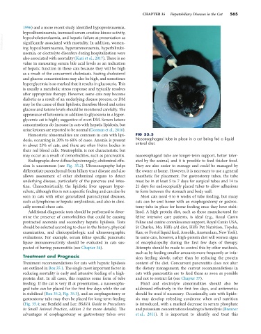
Hemostatic abnormalities are common in cats with lipi- FIG 35.3
dosis, occurring in 20% to 60% of cases. Anemia is present Nasoesophageal tube in place in a cat being fed a liquid
in about 25% of cats, and there are often Heinz bodies in enteral diet.
their red blood cells. Neutrophilia is not characteristic but
may occur as a result of comorbidities, such as pancreatitis. nasoesophageal tube are longer-term support, better toler-
Radiographs show diffuse hepatomegaly; abdominal effu- ated by the animal, and it is possible to feed thicker food.
sion is uncommon (see Fig. 35.2). Ultrasonography helps They are also easier to manage and could be managed by
differentiate parenchymal from biliary tract disease and also the owner at home. However, it is necessary to use a general
allows assessment of other abdominal organs to detect anesthetic for placement. For gastrostomy tubes, the tube
underlying disease, particularly of the pancreas and intes- must be in at least 5 to 7 days for surgical tubes and 14 to
tine. Characteristically, the lipidotic liver appears hyper- 21 days for endoscopically placed tubes to allow adhesions
echoic, although this is not a specific finding and can also be to form between the stomach and body wall.
seen in cats with other generalized parenchymal diseases, Most cats need 4 to 6 weeks of tube feeding, but many
such as lymphoma or hepatic amyloidosis, and also in clini- cats can be sent home with an esophagostomy or gastros-
cally normal obese cats. tomy tube in place for home feeding once they have stabi-
Additional diagnostic tests should be performed to deter- lized. A high-protein diet, such as those manufactured for
mine the presence of comorbidities that could be causing feline intensive care patients, is ideal (e.g., Royal Canin
protracted anorexia and secondary hepatic lipidosis. Tests Feline and canine convalescence support, Royal Canin USA,
should be selected according to clues in the history, physical St Charles, Mo; Hill’s a/d diet, Hill’s Pet Nutrition, Topeka,
examination, and clinicopathologic and ultrasonographic Kan; or Fortol liquid feed, Arnolds, Amsterdam, New York).
evaluations. For example, serum feline specific pancreatic In some cats, however, a high-protein diet will worsen signs
lipase immunoreactivity should be evaluated in cats sus- of encephalopathy during the first few days of therapy.
pected of having pancreatitis (see Chapter 34). Attempts should be made to control this by other methods,
such as by feeding smaller amounts more frequently or infu-
Treatment and Prognosis sion feeding slowly, rather than by reducing the protein
Treatment recommendations for cats with hepatic lipidosis content of the diet. Concurrent pancreatitis does not alter
are outlined in Box 35.1. The single most important factor in the dietary management; the current recommendations in
reducing mortality is early and intensive feeding of a high- cats with pancreatitis are to feed them as soon as possible
protein diet. In all cases, this requires some form of tube and not to restrict fat (see Chapter 37).
feeding. If the cat is very ill at presentation, a nasoesopha- Fluid and electrolyte abnormalities should also be
geal tube can be placed for the first few days while the cat addressed effectively in the first few days, and antiemetics
is stabilized (Box 35.2; Fig. 35.3), and an esophagostomy or should be used if necessary. Occasionally, cats with lipido-
gastrostomy tube may then be placed for long-term feeding sis may develop refeeding syndrome when oral nutrition
(Fig. 35.4; see Bexfield and Lee: BSAVA Guide to Procedures is introduced, with a marked decrease in serum phosphate
in Small Animal Practice, edition 2 for more details). The and potassium concentrations leading to hemolysis (Brenner
advantages of esophagostomy or gastrostomy tubes over et al., 2011). It is important to identify and treat this