
Page 595 - Small Animal Internal Medicine, 6th Edition
P. 595
CHAPTER 35 Hepatobiliary Diseases in the Cat 567
BOX 35.2 survival and that cats with secondary hepatic lipidosis may
do slightly worse than those with primary disease. However,
VetBooks.ir Placement of Nasoesophageal Feeding Tubes the differences were not significant, which suggests that it is
worth treating cats with secondary lipidosis as aggressively
This is used for short-term nutritional support (<1 wk) while
stabilizing the cat before the placement of an esophagos- as those with primary disease.
tomy or gastrostomy tube.
Placement BILIARY TRACT DISEASE
1. Premeasure tube to allow placement in caudal
esophagus, not stomach; this minimizes gastric reflux. Biliary tract diseases are the second most common disorders
Premeasure to seventh intercostal (IC) space from nose of the feline liver in the United States and the most common
or 75% of distance from nose to last rib if animal is feline liver disease in Europe (see Table 35.1). This contrasts
so obese that ribs cannot be counted (orogastric— with dogs, in which parenchymal diseases are most common.
ninth IC space or 90% of distance from nose to last All disorders of the biliary tract in cats can present with
rib). Mark tube with pen or piece of tape. similar clinical signs, including lethargy, anorexia, and jaun-
2. Apply local anesthetic to nose. Mild sedation may dice. Clinical, clinicopathologic, and diagnostic imaging
occasionally also be necessary, preferably with findings do not allow differentiation of the types of diseases;
buprenorphine or butorphanol. in most cases, cytology, bile culture, and histopathology of
3. Lubricate tube and advance into the ventral meatus; it
is important not to advance into the middle or dorsal the liver are necessary for accurate diagnosis and most effec-
meatus or the tube will lodge at the ethmoturbinates. It tive treatment as detailed in Table 35.3.
may be helpful to raise the cat’s head slightly to do
this. CHOLANGITIS
4. Hold the cat’s head normally as you approach the Cholangitis refers to inflammation of the biliary tract, which
pharynx to prevent tracheal intubation. Allow the cat in some (but not all) cats may also extend to the surrounding
to swallow, and advance the tube to a measured hepatic parenchyma (cholangiohepatitis). It is more common
mark or tape. in cats than in dogs, and it is typically divided into three
5. To check that the tube is correctly positioned, instill categories, likely associated with different etiologies—
water and air and auscultate over the left flank for neutrophilic cholangitis, lymphocytic cholangitis, and
bubbling in the stomach. If still uncertain, perform chronic cholangitis associated with liver fluke infestation.
radiography. If the tube does not have a radiodense
line, first inject some iodine-containing contrast The nomenclature of feline biliary tract disease has been
material into the tube. standardized by the World Small Animal Veterinary Asso-
6. Pass the tube over the top of the cat’s head, and ciation (WSAVA; Rothuizen et al., 2006; see Table 35.3).
suture or glue the tapes at the level of the nares and However, there is ongoing debate about the overlap between
top of the head; be careful to avoid interfering with lymphocytic cholangitis and chronic neutrophilic cholangi-
the cat’s whiskers. tis, and it has been suggested that these two categories should
7. Put on an Elizabethan collar. be combined into a broader non–suppurative cholangitis-
8. Flush regularly with warm water before and after cholangiohepatitis group (Warren et al., 2011). A wide
feeds. variety of alternative names have been used in the literature
in the past, sometimes blurring the categories and making
comparisons between studies difficult. It is likely that there
are several chronic forms of the disease with different etiolo-
gies and that increased understanding in the future will lead
to improved nomenclature (see Table 35.3).
Cats with inflammatory biliary tract disease also often
have concurrent pancreatitis and/or intestinal disease (often
termed triaditis, Fragkou et al., 2016). It has been proposed
that this is a reflection of the anatomy of their pancreatic and
bile ducts, which usually join before entering the proximal
duodenum through a common outflow tract. It has been
suggested that this increases the likelihood of intestinal con-
tents being refluxed up the pancreatic and bile ducts during
vomiting. It is also possible that sphincter of Oddi spasm
secondary to IBD, which has been reported in cats (Fur-
neaux, 2010), blocks the pancreatic and bile ducts in some
cats. However, it is likely that the reasons are multifactorial
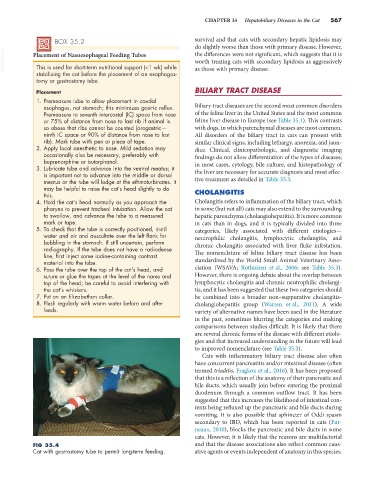
FIG 35.4 and that the disease associations also reflect common caus-
Cat with gastrostomy tube to permit long-term feeding. ative agents or events independent of anatomy in this species.