
Page 599 - Small Animal Internal Medicine, 6th Edition
P. 599
CHAPTER 35 Hepatobiliary Diseases in the Cat 571
K 1 SC or IM, q12h for 3 days) if there is any concern about a predilection for portal areas in cats, so it is an important
hemostasis; fresh-frozen plasma should be available to differential diagnosis in these cats. Large cell lymphomas are
VetBooks.ir manage potential postbiopsy bleeding. The author routinely relatively easy to diagnose, but small cell lymphomas appear
similar cytologically and histologically to lymphocytic chol-
administers vitamin K for 2 to 3 days before liver biopsy in
all cats. It is recommended that biopsies be taken from mul-
phocytic infiltrate extending beyond the limiting plate, an
tiple lobes because histologic findings can vary considerably angitis. Features suggesting lymphoma include a dense lym-
among liver lobes (Callahan Clark et al., 2011; Warren et al., absence of peribiliary fibrosis, and evidence of lymphoma in
2011). Bile aspiration is not necessary unless the disease is other tissues, such as the intestine and abdominal lymph
more acute and there is a possibility of neutrophilic cholan- nodes. The polymerase chain reaction (PCR) for antigen
gitis. Histology is important to rule out FIP (see Chapter 96) receptor rearrangement (PARR) assay (see Chapter 79) may
and lymphoma (see Chapter 79). Hepatic lymphoma shows be helpful in distinguishing lymphoma from inflammatory
disorders. The typical hepatic lesion in cats with FIP is a
multifocal pyogranulomatous reaction with evidence of vas-
culitis or perivasculitis, which is distinct from the periportal
lymphocytic infiltrate seen in cats with lymphocytic cholan-
gitis (Fig. 35.7). Serology or PCR assay for Bartonella spp.
might be considered, although the importance of this organ-
ism in the naturally occurring disease is unclear.
Treatment and Prognosis
Researchers disagree on the recommended therapy for this
disease, which likely reflects uncertainty about its etiology.
Several authors recommend immunosuppressive doses of
corticosteroids. However, although these tend to ameliorate
the acute flare-ups of the disease, they do not lead to resolu-
tion of signs, and the condition invariably recurs. Antibiotic
therapy is wise, at least early in treatment, until an infectious
FIG 35.6 etiology has been ruled out. There is a logical reason to use
Lateral abdominal radiograph from a cat with lymphocytic ursodeoxycholic acid (15 mg/kg PO q24h) in these cats for
cholangitis and associated ascites. The major differential
diagnosis in this case would be feline infectious peritonitis. its choleretic and antiinflammatory effects, as well as its
(Courtesy Diagnostic Imaging Department, Queen’s effect on modulating the bile acid pool and reducing toxic
Veterinary School Hospital, University of Cambridge, bile acids. Use of antioxidants such as S-adenosylmethionine
Cambridge, England.) (20 mg/kg or 200-400 mg total, once daily on an empty
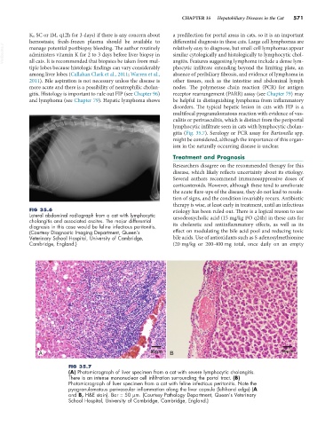
A 50 m BB 50 m
FIG 35.7
(A) Photomicrograph of liver specimen from a cat with severe lymphocytic cholangitis.
There is an intense mononuclear cell infiltration surrounding the portal tract. (B)
Photomicrograph of liver specimen from a cat with feline infectious peritonitis. Note the
pyogranulomatous perivascular inflammation along the liver capsule (left-hand edge) (A
and B, H&E stain). Bar = 50 µm. (Courtesy Pathology Department, Queen’s Veterinary
School Hospital, University of Cambridge, Cambridge, England.)