Page 590 - Small Animal Internal Medicine, 6th Edition
P. 590
562    PART IV   Hepatobiliary and Exocrine Pancreatic Disorders


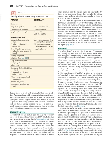
                   TABLE 35.1                                    obese animals, and the clinical signs are complicated by
                                                                 those of the concurrent disease. For example, the clinical
  VetBooks.ir  Clinically Relevant Hepatobiliary Diseases in Cats  signs of acute diabetic ketoacidosis are similar to those of
                                                                 developing hepatic lipidosis.
             PRIMARY
                                      SECONDARY
                                                                   Clinical signs are typical of an acute (reversible) loss of
                                                                 hepatocyte function and of hepatocyte swelling, with resul-
             Common                                              tant intrahepatic cholestasis. Cats are usually jaundiced and
             Idiopathic lipidosis     Secondary lipidosis        have intermittent vomiting and dehydration. They may also
             Neutrophilic cholangitis  Hyperthyroidism           have diarrhea or constipation. There is usually palpable hep-
             Lymphocytic cholangitis  Pancreatitis               atomegaly on physical examination. HE, most often mani-
                                      Diabetes mellitus          fested as depression and ptyalism, is related to severe
                                                                 hepatocellular dysfunction and relative arginine deficiency
             Uncommon or Rare                                    to which the anorexic cat is predisposed. Previously obese
             Congenital portosystemic   Secondary neoplasia (less   cats have extensive loss of muscle mass but maintain certain
               shunt                    common than primary)     fat stores, such as those in the falciform ligament and ingui-
             Extrahepatic bile duct   Biliary stasis associated   nal region (Fig. 35.2).
               obstruction              with extrahepatic sepsis
             Liver flukes (except common   Hepatic abscess       Diagnosis
               in hunting cats in endemic                        The only truly definitive and reliable method of diagnosing
               areas)                                            and identifying concurrent and causative conditions is his-
             Primary neoplasia                                   topathology of a wedge biopsy of liver obtained at laparot-
             Infections (see Box 35.5)                           omy or laparoscopy or (less reliably) a Tru-Cut-type biopsy
             Drug- or toxin-induced                              taken under ultrasonographic guidance. However, all of
               hepatopathy                                       these procedures require a general anesthetic, and most cats
             Biliary cysts                                       with hepatic lipidosis are too ill on presentation to be safely
                                                                 anesthetized. Therefore using fine-needle aspiration (FNA)
             Sclerosing cholangitis/biliary                      cytology of the liver performed blindly or under ultrasono-
               cirrhosis                                         graphic guidance in an awake or sedated cat can yield a
             Congenital ductal plate                             preliminary diagnosis; this will allow intensive management
               abnormalities                                     and tube feeding for a few days to stabilize the patient before
             Primary copper-associated                           anesthesia  is  considered  for  a  more  definitive  diagnosis.
               hepatopathy                                       Because coagulopathies are common in cats with lipidosis, a
             Hepatic amyloidosis                                 few days of therapy will help correct them before considering
             Intrahepatic arteriovenous                          surgery. The clinician must be aware, however, that FNA
               fistula                                           cytology, although useful for emergency diagnosis and man-
                                                                 agement, can be misleading in cats, and hepatic parenchymal
                                                                 disease can be misdiagnosed as lipidosis using this tech-
            disease and even in cats with a normal or thin body condi-  nique. In addition, concurrent diseases of the liver and other
            tion. Any anorexic cat with concurrent disease must there-  organs, including the pancreas and small intestine, will be
            fore be regarded as being at high risk of hepatic lipidosis, and   overlooked without a laparoscopic or surgical biopsy. It is
            appropriate feeding support should be instituted as rapidly   important to differentiate mild to moderate lipid accumula-
            as possible. Secondary lipidosis may occur in association   tion in hepatocytes, which is common in sick and anorexic
            with any disease causing anorexia, but has been most com-  cats and causes no clinical problems, from clinically severe
            monly recognized in cats with pancreatitis, diabetes mellitus   lipidosis on cytology (see Fig. 35.1).
            (DM), other hepatic disorders, IBD, and neoplasia.     FNA can be performed under ultrasonographic guidance
                                                                 while the cat is being evaluated or obtained blindly if there
            Clinical Features                                    is palpable hepatomegaly. The procedure is performed in a
            Most affected cats are middle-aged, but they can be of any   similar way  to  aspiration of  a  mass.  The enlarged  liver  is
            age or sex. In a recent study from Israel, 99% of affected cats   palpated, and the abdominal wall overlying it is clipped and
            were neutered and 66% were female (Kuzi et al., 2017). There   prepped. A 22-gauge needle is passed through the skin into
            is no reported breed predilection. Cats with primary lipido-  the liver from ventrally on the left side, which prevents inad-
            sis are commonly obese, are housed indoors, and have expe-  vertent puncturing of the gallbladder, and gentle suction is
            rienced a stressful event (e.g., introduction of a new pet into   applied to a 5-mL syringe two or three times, before with-
            the household, abrupt dietary change) or an illness that has   drawing and gently expressing the needle contents onto a
            caused them to become anorexic and lose weight rapidly. The   slide (see Fig. 34.14). Analgesia is recommended for either
            initiating event is not always known. Secondary lipidosis   procedure because puncture of the liver capsule is painful.
            may affect cats of normal or thin body condition as well as   Opiate partial agonists, such as buprenorphine, are a good

