
Page 586 - Small Animal Internal Medicine, 6th Edition
P. 586
558 PART IV Hepatobiliary and Exocrine Pancreatic Disorders
PANCREATIC BIOPSIES
Pancreatic biopsy is the only definitive way of making a
VetBooks.ir diagnosis but is not recommended in most cases. For any
biopsy to be justified clinically, the benefits should outweigh
the risks. Ultimately, the results should alter outcome in the
case, and this usually means they should alter treatment
decisions. In small animals, there are situations where a
biopsy might give prognostic rather than treatment informa-
tion and also alter outcome (for example, the owner may
decide on euthanasia as a result of finding a pancreatic
tumor). If the biopsy doesn’t do either of these things, then
it is difficult to justify ethically. It is difficult to biopsy a
pancreas noninvasively, and the procedure usually requires
laparotomy or laparoscopy. Fine needle aspiration cytology
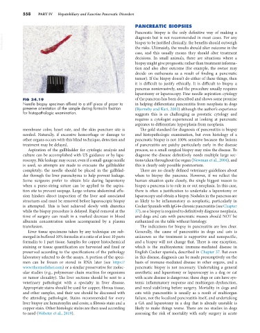
FIG 34.19 of the pancreas has been described and shows some promise
Needle biopsy specimen affixed to a stiff piece of paper to in helping differentiate pancreatitis from neoplasia in dogs
preserve orientation of the sample during formalin fixation (Bjorneby and Kari, 2002) although the author’s experience
for histopathologic examination. suggests this is as challenging as prostatic cytology and
requires a cytologist experienced at looking at pancreatic
aspirates to differentiate hyperplasia from neoplasia.
membrane color, heart rate, and the skin puncture site is The gold standard for diagnosis of pancreatitis is biopsy
needed. Naturally, if excessive hemorrhage or damage to and histopathologic examination, but even histology of a
other organs occurs with this blind technique, detection and pancreatic biopsy is not 100% sensitive because the lesions
treatment may be delayed. of pancreatitis are patchy particularly early in the disease
Aspiration of the gallbladder for cytologic analysis and process, so a small surgical biopsy may miss the disease. To
culture can be accomplished with US guidance or by lapa- diagnose the disease definitively needs multiple large sec-
roscopy. Bile leakage may occur, even if a small-gauge needle tions taken throughout the organ (Newman et al., 2004), and
is used, so attempts are made to evacuate the gallbladder this is clearly only possible postmortem.
completely; the needle should be placed in the gallblad- There are no clearly defined veterinary guidelines about
der through the liver parenchyma to help prevent leakage. when to biopsy the pancreas. However, if we reflect the
Some surgeons prefer to obtain bile during laparotomy human situation quite closely, the single biggest reason to
when a purse-string suture can be applied to the aspira- biopsy a pancreas is to rule in or out neoplasia. In this case,
tion site to prevent seepage. Large-volume abdominal effu- there is often a justification to undertake a laparotomy or
sion hinders direct inspection of the liver and associated laparoscopy and obtain a biopsy. Nodules in the pancreas are
structures and must be removed before laparoscopic biopsy as likely to be inflammatory as neoplastic, particularly in
is attempted. This is best achieved slowly with diuretics Cocker Spaniels with IgG4+ chronic pancreatitis (see Chapter
while the biopsy procedure is delayed. Rapid removal at the 37), so a biopsy is required to definitively diagnose neoplasia,
time of surgery can result in a marked decrease in blood and dogs and cats with pancreatic masses should NOT be
albumin concentration unless accompanied by a plasma euthanized on the table without histology.
transfusion. The indications for biopsy in pancreatitis are less clear.
Liver tissue specimens taken by any technique are sub- Generally, the cause of pancreatitis in dogs and cats is
merged in buffered 10% formalin at a ratio of at least 10 parts unknown so the treatment is supportive and nonspecific,
formalin to 1 part tissue. Samples for copper histochemical and a biopsy will not change that. There is one exception,
staining or tissue quantification are harvested and fixed or which is the multisystemic immune-mediated disease in
preserved according to the specifications of the pathology English Cocker spaniels, described in Chapter 37. But even
laboratory selected to do the assays. A portion of the speci- in this disease, diagnosis can be made presumptively on the
men can be frozen or stored in RNA later (see https:// basis of immune-mediated disease in other organs, and a
www.thermofisher.com) or a similar preservative for molec- pancreatic biopsy is not necessary. Undertaking a general
ular studies (e.g., polymerase chain reaction for organisms anesthetic and laparotomy or laparoscopy in a dog or cat
or tumor clonality). The liver sections should be sent to a with acute disease is dangerous; these dogs or cats have sys-
veterinary pathologist with a specialty in liver disease. temic inflammatory response and multiorgan dysfunction,
Appropriate stains should be used for copper, fibrous tissue, and need stabilizing before surgery. Mortality in dogs and
and other samples, and their use should be discussed with cats with pancreatitis is usually as a result of multiorgan
the attending pathologist. Stains recommended for every failure, not the localized pancreatitis itself, and undertaking
liver biopsy are hematoxylin and eosin; a fibrosis stain and a a GA and laparotomy in a dog that is already unstable is
copper stain. Other histologic stains are then used according likely to make things worse. There are no studies in dogs
to need (Webster et al., 2019). assessing the risk of mortality with early surgery in acute