
Page 581 - Small Animal Internal Medicine, 6th Edition
P. 581
CHAPTER 34 Diagnostic Tests for the Hepatobiliary and Pancreatic System 553
follows that acute cases of pancreatitis, which classically With acute pancreatitis, dogs had an enlarged, homogenously
show these features, are more often visible ultrasonographi- to heterogeneously attenuating and contrast-enhancing pan-
VetBooks.ir cally than low grade, chronic forms with little edema. US is creas with ill-defined borders, as has been reported in
humans (Adrian et al., 2015).
also useful to assess for concurrent pancreatic masses,
abscesses, or pseudocysts and to detect associated cholangi-
tis and small intestinal wall thickening. An animal with acute MAGNETIC RESONANCE IMAGING
pancreatitis usually shows a characteristically focal area of MRI is used frequently in human medicine for imaging of
pain as the ultrasound probe is positioned over the the biliary tract and pancreatic ducts. MR cholangiopancrea-
pancreas. tography allows accurate imaging of duct abnormalities
without any need for contrast. There are no reports yet of its
COMPUTED TOMOGRAPHY clinical use in dogs and cats, but a recent study in normal
CT is increasingly available in veterinary medicine and can cats showed promise for future clinical use (Marolf et al.,
be used to image a variety of hepatic diseases and masses. It 2011; Marolf 2017).
is most commonly indicated in imaging PSS and has now
largely replaced contrast radiography in identifying PSS and SCINTIGRAPHY
giving detailed anatomic information (Nelson and Nelson, Scintigraphy is used in hepatobiliary disease in cats and dogs
2011). CT requires a general anesthetic but is less invasive most frequently to diagnose PSS. The isotope selected is
than contrast radiography. US can be performed under seda- technetium-99m ( 99m Tc). The isotope has a short half-life (6
tion and is less expensive than CT. Therefore, if a shunt can hours); thus although the animal must be relatively isolated
be accurately identified with plain or bubble US, a CT is not for 24 to 48 hours, and urinary and fecal waste stored until
necessarily indicated. However, with complex shunts of radioactivity has fallen to background levels, there is minimal
unclear anatomy on US, CT can provide invaluable informa- radiation hazard to the animal or involved personnel. For
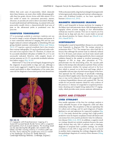
tion before surgery (Fig. 34.13). diagnosis of PSS in dogs, after placement of 99m Tc-
Abdominal CT has so far proved largely disappointing for pertechnetate into the descending colon, the vascular path
the diagnosis of pancreatitis in dogs and cats, although a taken by the isotope after absorption is plotted. Time-activity
recent study suggested it might be more reliable than ultra- curves determine whether the isotope arrived in the liver
sound, particularly to allow visualization of the entire pan- first, which is normal, or in the heart and lungs, which is
creas and for diagnosis of associated portal vein thrombosis. compatible with any type of portal venous bypass of the liver.
This approach has the advantage of specifically evaluating
the portal blood supply rather than the hepatic mass, which
may or may not be reduced in animals with congenital PSS
or primary hepatobiliary disease and acquired PSS. The test
results do not provide anatomic detail but only evidence of
the presence or absence of congenital or acquired portosys-
temic shunting and is largely being replaced by CT angiog-
raphy, which provides more precise anatomic information.
BIOPSY AND CYTOLOGY
LIVER CYTOLOGY
Fine-needle aspiration of the liver for cytologic analysis is
rarely advisable because of low diagnostic yield and often
misleading results. The exceptions to this are aspirating bile
for culture and cytology, and for quick diagnosis of hepatic
lipidosis in cats and aspiration for suspected neoplasia (Fig.
34.14). One study showed that cytology had a high positive
predictive value for hepatic neoplasia (round cell tumors and
carcinoma) but a low positive predictive value for inflamma-
FIG 34.13 tory disease (Bahr et al., 2013). In addition, a negative result
CT angiogram with three-dimensional mapping of a on cytology should not exclude neoplasia because the same
2-year-old neutered female Border Terrier with a congenital study showed that only 50% of patients with a histologic
portosystemic shunt of unusual anatomy. The shunting vessel diagnosis of neoplasia had a positive cytology. An overall
can be seen leaving the left gastric vein cranial to the
kidney and then coursing cranially in a tortuous path over correlation of only 30% in dogs and 51% in cats was found
the liver, finally entering the caudal vena cava just caudal in another study comparing the cytologic diagnosis with the
to the diaphragm. (Courtesy Dr. Paddy Mannion, histopathologic diagnosis of a variety of liver diseases (Wang
Cambridge Radiology Referrals, Cambridge, England.) et al., 2004).