
Page 582 - Small Animal Internal Medicine, 6th Edition
P. 582
554 PART IV Hepatobiliary and Exocrine Pancreatic Disorders
BOX 34.4
VetBooks.ir Patient and Operator Considerations for Hepatic Biopsy
Patient
1. Characteristics of the suspected hepatobiliary
disorder—liver size (small, normal, enlarged); texture
(fibrotic or friable); focal, multifocal, or diffuse
distribution; presence of abdominal effusion
2. Clinical stability and suitability for anesthesia
3. Coagulation status and platelet count
Operator
1. Available equipment
2. Experience with chosen technique
3. Complication rate for chosen technique
4. Size of specimen needed
5. Access to reliable veterinary pathology laboratory
6. Cost of procedure and client finances
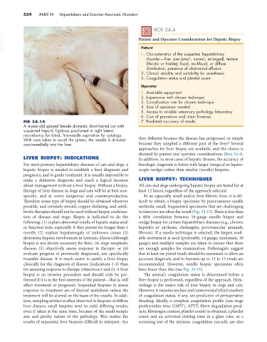
FIG 34.14 7. Predicted accuracy of results
A 4-year-old spayed female domestic short-haired cat with
suspected hepatic lipidosis positioned in right lateral
recumbency for blind, fine-needle aspiration for cytology.
With care taken to avoid the spleen, the needle is directed they different because the disease has progressed or simply
craniomedially into the liver. because they sampled a different part of the liver? Several
approaches for liver biopsy are available, and the choice is
dictated by patient and operator considerations (Box 34.4).
LIVER BIOPSY: INDICATIONS In addition, in most cases of hepatic disease, the accuracy of
For most primary hepatobiliary diseases of cats and dogs, a histologic diagnosis is better with larger (surgical or laparo-
hepatic biopsy is needed to establish a final diagnosis and scopic wedge) rather than smaller (needle) biopsies.
prognosis, and to guide treatment. It is usually impossible to
make a definitive diagnosis and reach a logical decision LIVER BIOPSY: TECHNIQUES
about management without a liver biopsy. Without a biopsy, All cats and dogs undergoing hepatic biopsy are fasted for at
therapy of liver disease in dogs and cats will be at best non- least 12 hours, regardless of the approach selected.
specific, and at worst dangerous and counterproductive. In an especially small and/or firm fibrotic liver, it is dif-
Therefore some type of biopsy should be obtained wherever ficult to obtain a biopsy specimen by percutaneous needle
possible, and certainly steroid, copper-chelating, and antifi- methods; small, fragmented specimens that are challenging
brotic therapies should not be used without biopsy confirma- to interpret are often the result (Fig. 34.15). There is less than
tion of disease and stage. Biopsy is indicated to do the a 40% correlation between 18-gauge needle biopsy and
following: (1) explain abnormal results of hepatic status and/ wedge biopsy for certain hepatobiliary diseases (e.g., chronic
or function tests, especially if they persist for longer than 1 hepatitis or cirrhosis, cholangitis, portovascular anomaly,
month; (2) explain hepatomegaly of unknown cause; (3) fibrosis). If a needle technique is selected, the largest avail-
determine hepatic involvement in systemic illness (although able instrument is used (preferably, 14 gauge; minimum, 16
biopsy is not always necessary for this); (4) stage neoplastic gauge) and multiple samples are taken to ensure that there
disease; (5) objectively assess response to therapy; or (6) are enough samples for examination. Pathologists suggest
evaluate progress of previously diagnosed, not specifically that at least six portal triads should be examined to allow an
treatable disease. It is much easier to justify a liver biopsy accurate diagnosis, and in humans up to 12 to 15 triads are
clinically for the diagnosis of disease (indications 1-4) than recommended. However, needle biopsy specimens often
for assessing response to therapy (objectives 5 and 6). A liver have fewer than this (see Fig. 34.15).
biopsy is an invasive procedure and should only be per- The animal’s coagulation status is determined before a
formed if it is in the best interests of the patient—that is, will liver biopsy is performed, regardless of the approach. Hem-
affect treatment or prognosis. Sequential biopsies to assess orrhage is the major risk of liver biopsy in dogs and cats.
response to treatment are of limited usefulness unless the However, it remains unclear and controversial which markers
treatment will be altered on the basis of the results. In addi- of coagulation status, if any, are predictive of perioperative
tion, sampling artifact is often observed in biopsies of diffuse bleeding. Ideally, a complete coagulation profile (one-stage
liver disease; small biopsies tend to yield differing results, prothrombin time [OSPT], APTT, fibrin degradation prod-
even if taken at the same time, because of the small sample ucts, fibrinogen content, platelet count) is obtained; a platelet
size and patchy nature of the pathology. This makes the count and an activated clotting time in a glass tube, as a
results of sequential liver biopsies difficult to interpret. Are screening test of the intrinsic coagulation cascade, are also