
Page 577 - Small Animal Internal Medicine, 6th Edition
P. 577
CHAPTER 34 Diagnostic Tests for the Hepatobiliary and Pancreatic System 549
VetBooks.ir
FIG 34.8
Lateral abdominal radiograph from a 7-year-old Jack Russell FIG 34.9
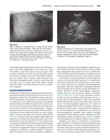
Terrier with acute pancreatitis. There are minimal changes Abdominal ultrasound is enhanced by the presence of
apparent apart from a mild loss of abdominal contrast, in ascites. Shown is an ultrasound of the abdomen of a dog
spite of the severity of the disease. This does, however, help with chronic hepatitis and ascites. (Courtesy Diagnostic
rule out acute obstruction because the intestinal loops are Imaging Department, Queen’s Veterinary School Hospital,
not dilated and gas-filled. (Courtesy Diagnostic Imaging University of Cambridge, Cambridge, England.)
Department, Queen’s Veterinary School Hospital, University
of Cambridge, Cambridge, England.)
and laterally displaced proximal duodenum on ventrodorsal large hepatic and portal veins, and adjacent caudal vena cava
views; and caudal displacement of the transverse colon. are all visible in the liver of the normal cat and dog. Unlike
Occasionally, a mass effect may be seen in the region of the plain radiography, which requires two views to complete the
pancreas, usually the result of fat necrosis. Pancreatic tumors study, US makes many slices through several planes to create
by contrast are usually small, but it is impossible to differen- a three-dimensional reconstruction of the target structures.
tiate fat necrosis from neoplasia using imaging alone. Performing US and interpreting the recorded images are
Abdominal radiographs appear normal in many dogs and a blend of technical skill and experience. It is important to
cats with acute or chronic pancreatitis. Barium studies note that the sensitivity of US in hepatic disease is not 100%.
should be avoided, if possible, because they do not contrib- In a recent study, the liver appeared ultrasonographically
ute to diagnosis. abnormal in only 48% of dogs with histologically confirmed
chronic hepatitis and in only 68% of dogs with hepatic lym-
ULTRASONOGRAPHY phoma. Therefore a normal ultrasonographic appearance
Abdominal US is the preferred diagnostic modality for evalu- certainly does not rule out hepatic disease or neoplasia
ating the hepatobiliary system in dogs and cats. However, it is (Warren-Smith et al., 2012). It is also important to remem-
important to realize that it has limited sensitivity and speci- ber that US does not diagnose what the lesions are (i.e., it
ficity for liver disease. Operating on the principle that a pulse cannot yield a histologic diagnosis). With a few exceptions,
of sound (echo) can be reflected when it passes through the which predominantly involve lesions of the biliary tract and
interface between two different materials, US can detect dif- vessels, the ultrasonographic appearance of a variety of
ferences between homogeneous liquids of low echogenicity, benign and malignant hepatic lesions can appear similar and
such as blood and bile, and more heterogeneous echogenic histology of a liver biopsy is usually required for diagnosis.
structures made up of several soft tissues. Whereas abdomi- An animal should never be euthanized on the basis of an
nal effusion obscures abdominal detail on survey radiog- ultrasonographically identified tumor without histologic
raphy, it enhances the ability of US to detect abnormalities confirmation because benign nodular hyperplasia or focal
(Fig. 34.9). However, bone and gas-filled organs reflect the inflammatory lesions can look the same. Table 34.5 outlines
sound beam completely (acoustic shadowing), so that struc- the typical appearances of different hepatic lesions on US.
tures beneath cannot be imaged by US. The procedure does Neoplasia may appear as hyperechoic or hypoechoic
not require anesthesia but the patient must be still, and good and focal, diffuse, or normal liver. Hepatic lymphoma often
contact between the transducer and abdominal skin must appears diffusely hypoechoic but can also appear hyperechoic
be ensured by clipping the haircoat and applying acoustic or normal. Some tumors, such as metastatic hemangiosarco-
coupling gel. Animals are usually positioned in dorsal or mas, have a classically nodular hypoechoic appearance (Fig.
lateral recumbency. The hepatic parenchyma, gallbladder, 34.10) and may appear as target lesions, which are relatively