
Page 575 - Small Animal Internal Medicine, 6th Edition
P. 575
CHAPTER 34 Diagnostic Tests for the Hepatobiliary and Pancreatic System 547
VetBooks.ir
A B
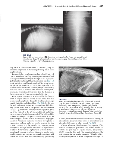
FIG 34.4
Lateral (A) and ventrodorsal (B) abdominal radiographs of a 9-year-old spayed female
mixed-breed dog with a hepatocellular carcinoma enlarging the right lateral liver lobe.
The dog was also severely hypoglycemic.
may result in caudal displacement of the liver, giving the
erroneous impression of hepatomegaly using other radio-
graphic criteria.
Because the liver may be contained entirely within the rib
cage in normal cats and dogs, microhepatia is more difficult
to recognize than hepatomegaly. Changes in the angle of the
gastric fundus in the right lateral projection (see Fig. 34.2)
could indicate a small hepatic shadow if the angle is more
upright or perpendicular to the spine, especially if the
stomach seems rather close to the diaphragm. The liver may
also seem small in animals with traumatic diaphragmatic
hernia and herniation of liver lobes into the thorax or in
those with congenital peritoneopericardial hernia.
Focal hepatic enlargement is indicated by the displace-
ment of organs adjacent to the affected lobe. The most FIG 34.5
common radiographically detectable focal hepatic enlarge- Lateral abdominal radiograph of a 12-year-old neutered
ment is that of the right lateral lobe (Fig. 34.4). In this case, male domestic short-haired cat with chronic cholangitis,
the body and pyloric regions of the stomach are shifted dor- cholecystitis, and pancreatitis. Note the radiodensity
sally (lateral view) and to the patient’s left (ventrodorsal overlying the liver shadow, which was identified at surgery
view); the gastric fundus remains in normal position. Shift- as a gallstone in the common bile duct. (Courtesy
ing of the stomach to the left is normal in cats and should Diagnostic Imaging Department, Queen’s Veterinary School
Hospital, University of Cambridge, Cambridge, England.)
not be mistaken for right hepatomegaly. If the left lateral lobe
or lobes are enlarged, the gastric fundus moves to the left
and caudally; the lesser curvature of the stomach may appear bacteria (patchy and/or linear areas of decreased opacity) or
indented. Primary or metastatic neoplasia, hyperplastic or mineralization (focal or diffuse spots of mineralization or
regenerative nodules, and cysts usually account for focal mineralized bile ducts or biliary calculi; Fig. 34.5).
hepatic enlargement or for irregular liver margins without With the advent of US and computed tomography (CT),
enlargement. If the gallbladder is massively enlarged because contrast radiographic procedures are seldom needed to
of EBDO, it may mimic a right cranial abdominal mass or confirm the presence of hepatic masses, cholelithiasis,
an enlarged, rounded liver lobe. Changes in hepatic radio- EBDO, congenital PSS, and other structural diseases. The
graphic opacity are rare and are usually associated with contrast study that can localize congenital PSS and is achiev-
hepatic or biliary tract infection caused by gas-forming able in private practice is portal venography, although CT