
Page 574 - Small Animal Internal Medicine, 6th Edition
P. 574
546 PART IV Hepatobiliary and Exocrine Pancreatic Disorders
of the liver (see Table 33.1). Optimally, the animal should breeds with narrow, deep chests, the entire liver shadow may
have an empty GI tract when the radiographs are obtained. be contained within the caudal rib cage. In dogs with a wide,
VetBooks.ir In the normal dog and cat in right lateral recumbency, the shallow thoracic conformation, the liver may extend slightly
beyond the costal arch. In the ventrodorsal view, the borders
gastric axis is parallel to the ribs at the 10th intercostal space,
and the caudoventral border of the liver (the left lateral liver
gastric fundus; in this view, the gastric shadow is perpen-
lobe) appears sharp. The image is made possible by the con- of the liver are defined by the cranial duodenum and the
trasting fat-filled falciform ligament (Fig. 34.2). In dog dicular to the spine. This view is less useful for assessing liver
size unless it is markedly and asymmetrically enlarged. The
gallbladder and extrahepatic biliary tree are not separately
visible radiographically in healthy animals.
Survey radiography is of minimal to no benefit if there is
moderate to marked abdominal effusion because the similar
radiographic opacities of the liver and fluid preclude distinc-
tion of liver size and shape, except by indirect assessment
(e.g., malposition of a gas-filled stomach and duodenum;
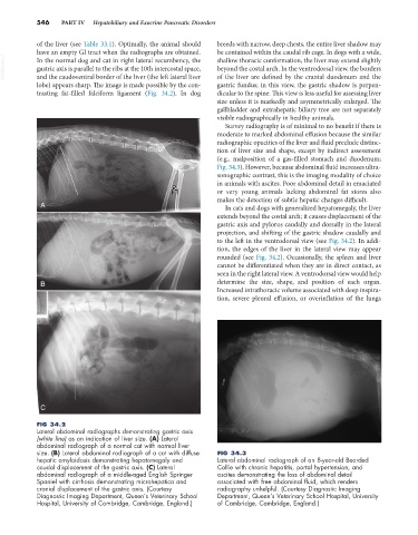
Fig. 34.3). However, because abdominal fluid increases ultra-
sonographic contrast, this is the imaging modality of choice
in animals with ascites. Poor abdominal detail in emaciated
or very young animals lacking abdominal fat stores also
makes the detection of subtle hepatic changes difficult.
A In cats and dogs with generalized hepatomegaly, the liver
extends beyond the costal arch; it causes displacement of the
gastric axis and pylorus caudally and dorsally in the lateral
projection, and shifting of the gastric shadow caudally and
to the left in the ventrodorsal view (see Fig. 34.2). In addi-
tion, the edges of the liver in the lateral view may appear
rounded (see Fig. 34.2). Occasionally, the spleen and liver
cannot be differentiated when they are in direct contact, as
seen in the right lateral view. A ventrodorsal view would help
B determine the size, shape, and position of each organ.
Increased intrathoracic volume associated with deep inspira-
tion, severe pleural effusion, or overinflation of the lungs
C
FIG 34.2
Lateral abdominal radiographs demonstrating gastric axis
(white line) as an indication of liver size. (A) Lateral
abdominal radiograph of a normal cat with normal liver
size. (B) Lateral abdominal radiograph of a cat with diffuse FIG 34.3
hepatic amyloidosis demonstrating hepatomegaly and Lateral abdominal radiograph of an 8-year-old Bearded
caudal displacement of the gastric axis. (C) Lateral Collie with chronic hepatitis, portal hypertension, and
abdominal radiograph of a middle-aged English Springer ascites demonstrating the loss of abdominal detail
Spaniel with cirrhosis demonstrating microhepatica and associated with free abdominal fluid, which renders
cranial displacement of the gastric axis. (Courtesy radiography unhelpful. (Courtesy Diagnostic Imaging
Diagnostic Imaging Department, Queen’s Veterinary School Department, Queen’s Veterinary School Hospital, University
Hospital, University of Cambridge, Cambridge, England.) of Cambridge, Cambridge, England.)