
Page 576 - Small Animal Internal Medicine, 6th Edition
P. 576
548 PART IV Hepatobiliary and Exocrine Pancreatic Disorders
angiography is the preferred method to diagnose PSS when after congenital PSS ligation if there is concern about the
available. Acceptable approaches for portal venography are adequacy of the intrahepatic portal vasculature. In addition,
VetBooks.ir splenoportography, operative mesenteric portography, and it has been shown that the degree of intrahepatic portal
vessel opacification on postligation portography is predictive
operative splenoportography. The two operative procedures
require general anesthesia and a small abdominal incision;
Abdominal radiographs in patients with pancreatitis
however, little sophisticated equipment is needed, and these for outcome (Lee et al., 2006).
procedures are associated with few complications. usually show mild or no changes, even in those with severe
A 22-gauge catheter is placed in the splenic vein or a disease (Fig. 34.8). However, in patients with acute disease,
mesenteric vein (Fig. 34.6), and the resting portal venous abdominal radiography plays an important role in ruling out
pressure is measured with a water manometer (normal = acute intestinal obstruction, which would result in obvious
6-13 cm H 2 O). Portal pressure is measured as soon as pos- changes, primarily dilated, gas-filled, stacking loops of intes-
sible in the procedure because prolonged anesthesia may tine and the presence of radiopaque foreign bodies. Typical
complicate its interpretation. An injection of iodine-based radiographic changes in dogs and cats with acute pancreatitis
contrast medium, 0.5 to 1 mL/kg, is then quickly made. include a focal decrease in contrast in the cranial abdomen
Lateral and possibly ventrodorsal and oblique radiographs associated with local peritonitis; a dilated, fixed (C-shaped),
are made at the end of the injection. Contrast medium given
to a normal cat or dog should flow into the portal vein, enter
the liver, and branch multiple times, opacifying the extrahe-
patic and intrahepatic portal vasculature. Diversion of the
contrast medium into the systemic circulation indicates PSS
(Fig. 34.7). Measurement of portal pressure and a liver biopsy
can be performed during the operative techniques; they are
required to distinguish acquired PSS from congenital PSS,
which is essential to rendering an accurate prognosis and
developing the correct treatment plan. As a general rule,
cases of congenital PSS are usually single, whereas acquired
PSS are multiple, so the mesenteric portography may suggest
a diagnosis. It may be necessary to repeat the contrast study
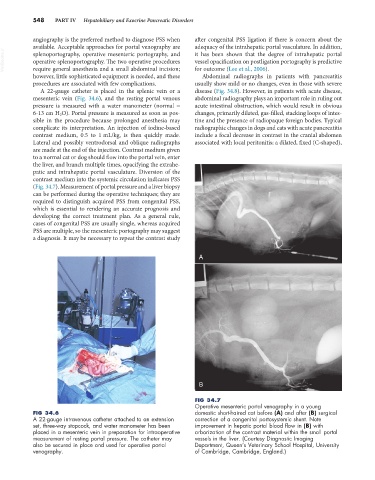
A
B
FIG 34.7
Operative mesenteric portal venography in a young
FIG 34.6 domestic short-haired cat before (A) and after (B) surgical
A 22-gauge intravenous catheter attached to an extension correction of a congenital portosystemic shunt. Note
set, three-way stopcock, and water manometer has been improvement in hepatic portal blood flow in (B) with
placed in a mesenteric vein in preparation for intraoperative arborization of the contrast material within the small portal
measurement of resting portal pressure. The catheter may vessels in the liver. (Courtesy Diagnostic Imaging
also be secured in place and used for operative portal Department, Queen’s Veterinary School Hospital, University
venography. of Cambridge, Cambridge, England.)