
Page 580 - Small Animal Internal Medicine, 6th Edition
P. 580
552 PART IV Hepatobiliary and Exocrine Pancreatic Disorders
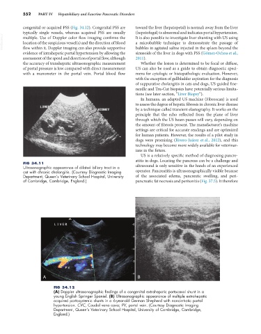
congenital or acquired PSS (Fig. 34.12). Congenital PSS are toward the liver (hepatopetal) is normal; away from the liver
typically single vessels, whereas acquired PSS are usually (hepatofugal) is abnormal and indicates portal hypertension.
VetBooks.ir multiple. Use of Doppler color flow imaging confirms the It is also possible to investigate liver shunting with US using
a microbubble technique to demonstrate the passage of
location of the suspicious vessel(s) and the direction of blood
flow within it. Doppler imaging can also provide supportive
sinusoids of the liver in dogs with PSS (Gómez-Ochoa et al.,
evidence of intrahepatic portal hypertension by allowing the bubbles in agitated saline injected in the spleen beyond the
assessment of the speed and direction of portal flow, although 2011).
the accuracy of transhepatic ultrasonographic measurement Whether the lesion is determined to be focal or diffuse,
of portal pressure is low compared with direct measurement US can also be used as a guide to obtain diagnostic speci-
with a manometer in the portal vein. Portal blood flow mens for cytologic or histopathologic evaluation. However,
with the exception of gallbladder aspiration for the diagnosis
of suppurative cholangitis in cats and dogs, US-guided fine-
needle and Tru-Cut biopsies have potentially serious limita-
tions (see later section, “Liver Biopsy”).
In humans, an adapted US machine (Fibroscan) is used
to assess the degree of hepatic fibrosis in chronic liver disease
by a technique called transient elastography. It works on the
principle that the echo reflected from the plane of liver
through which the US beam passes will vary, depending on
the amount of fibrosis present. The manufacturer’s machine
settings are critical for accurate readings and are optimized
for human patients. However, the results of a pilot study in
dogs were promising (Rivero-Juárez et al., 2012), and this
technology may become more widely available for veterinar-
ians in the future.
US is a relatively specific method of diagnosing pancre-
atitis in dogs. Locating the pancreas can be a challenge and
FIG 34.11
Ultrasonographic appearance of dilated biliary tract in a ultrasound is only sensitive in the hands of an experienced
cat with chronic cholangitis. (Courtesy Diagnostic Imaging operator. Pancreatitis is ultrasonographically visible because
Department, Queen’s Veterinary School Hospital, University of the associated edema, pancreatic swelling, and peri-
of Cambridge, Cambridge, England.) pancreatic fat necrosis and peritonitis (Fig. 37.5). It therefore
A B
FIG 34.12
(A) Doppler ultrasonographic findings of a congenital extrahepatic portocaval shunt in a
young English Springer Spaniel. (B) Ultrasonographic appearance of multiple extrahepatic
acquired portosystemic shunts in a 6-year-old German Shepherd with noncirrhotic portal
hypertension. CVC, Caudal vena cava; PV, portal vein. (Courtesy Diagnostic Imaging
Department, Queen’s Veterinary School Hospital, University of Cambridge, Cambridge,
England.)