
Page 584 - Small Animal Internal Medicine, 6th Edition
P. 584
556 PART IV Hepatobiliary and Exocrine Pancreatic Disorders
be taken under heavy sedation or general anesthesia, and et al., 2016). Recovery is much quicker than with laparot-
are better than no biopsy at all. However, they are often omy, and animals can usually be sent home on the same day
VetBooks.ir too small and nonrepresentative (see Fig. 34.15), and it is as the procedure. With laparotomy and laparoscopy, consid-
eration should be given to obtaining a bile sample by aspira-
not possible to obtain enough tissue for quantitative copper
measurement by Tru-Cut needle biopsy. Multiple biopsies
such as the pancreas at the same time, as indicated in the
should be taken with as large a Tru-Cut needle as possible to tion and examining and taking biopsies from other organs
maximize the chances of obtaining diagnostic samples. The
animal should be monitored carefully for hemorrhage after-
ward (preferably hospitalized overnight), which although
uncommon, can develop unnoticed in these animals and
can be life-threatening.
Laparotomy is much more invasive but allows examina-
tion of other abdominal organs (e.g., pancreas, small intes-
tine), observation of the liver, and careful biopsy. The risk of
hemorrhage is, therefore smaller than with Tru-Cut biopsies
because any bleeding can be seen and dealt with at the time
of surgery. The biopsies obtained are generally bigger and
more diagnostic except that focal lesions deep in the paren-
chyma may be missed if US has not been used as well. If one
part of the liver looks normal and another part abnormal,
biopsies should be obtained from both areas because the
apparently normal part may be the diseased area. Laparot-
omy is clearly indicated if there is a mass that could be
resected (Fig. 34.16). Laparoscopy is less invasive than lapa-
rotomy and has similar advantages. It is the preferred method FIG 34.16
Laparotomy for resection of a hepatic mass in a dog. A
of obtaining diagnostic liver biopsies if there is access to diagnosis of hepatocellular carcinoma was made on
equipment and operator experience (Fig. 34.17), and has histopathology. (Courtesy Dr. Laura Owen, Soft Tissue
been shown to have a low complication rate, even in dogs Surgery Department, Queen’s Veterinary School Hospital,
with advanced liver disease and coagulopathies (McDevitt University of Cambridge, Cambridge, England.)
A B
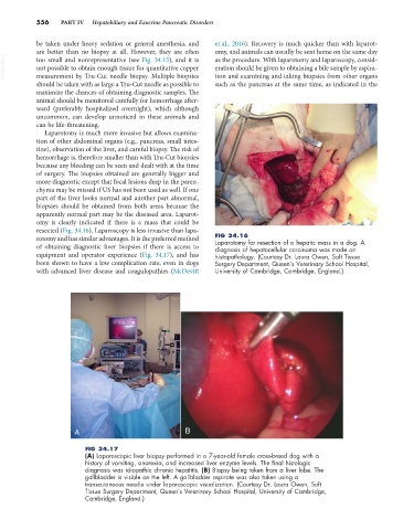
FIG 34.17
(A) Laparoscopic liver biopsy performed in a 7-year-old female cross-breed dog with a
history of vomiting, anorexia, and increased liver enzyme levels. The final histologic
diagnosis was idiopathic chronic hepatitis. (B) Biopsy being taken from a liver lobe. The
gallbladder is visible on the left. A gallbladder aspirate was also taken using a
transcutaneous needle under laparoscopic visualization. (Courtesy Dr. Laura Owen, Soft
Tissue Surgery Department, Queen’s Veterinary School Hospital, University of Cambridge,
Cambridge, England.)