Page 578 - Small Animal Internal Medicine, 6th Edition
P. 578

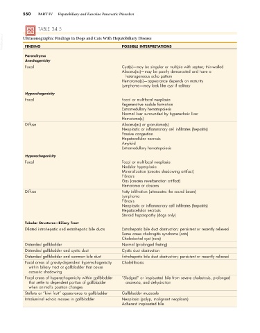
550    PART IV   Hepatobiliary and Exocrine Pancreatic Disorders


                   TABLE 34.5
  VetBooks.ir  Ultrasonographic Findings in Dogs and Cats With Hepatobiliary Disease

                                                          POSSIBLE INTERPRETATIONS
             FINDING
             Parenchyma
             Anechogenicity
             Focal                                        Cyst(s)—may be singular or multiple with septae; thin-walled
                                                          Abscess(es)—may be poorly demarcated and have a
                                                            heterogeneous echo pattern
                                                          Hematoma(s)—appearance depends on maturity
                                                          Lymphoma—may look like cyst if solitary

             Hypoechogenicity
             Focal                                        Focal or multifocal neoplasia
                                                          Regenerative nodule formation
                                                          Extramedullary hematopoiesis
                                                          Normal liver surrounded by hyperechoic liver
                                                          Hematoma(s)
             Diffuse                                      Abscess(es) or granuloma(s)
                                                          Neoplastic or inflammatory cell infiltrates (hepatitis)
                                                          Passive congestion
                                                          Hepatocellular necrosis
                                                          Amyloid
                                                          Extramedullary hematopoiesis
             Hyperechogenicity
             Focal                                        Focal or multifocal neoplasia
                                                          Nodular hyperplasia
                                                          Mineralization (creates shadowing artifact)
                                                          Fibrosis
                                                          Gas (creates reverberation artifact)
                                                          Hematoma or abscess
             Diffuse                                      Fatty infiltration (attenuates the sound beam)
                                                          Lymphoma
                                                          Fibrosis
                                                          Neoplastic or inflammatory cell infiltrates (hepatitis)
                                                          Hepatocellular necrosis
                                                          Steroid hepatopathy (dogs only)

             Tubular Structures—Biliary Tract
             Dilated intrahepatic and extrahepatic bile ducts  Extrahepatic bile duct obstruction; persistent or recently relieved
                                                          Some cases cholangitis syndrome (cats)
                                                          Choledochal cyst (rare)
             Distended gallbladder                        Normal (prolonged fasting)
             Distended gallbladder and cystic duct        Cystic duct obstruction
             Distended gallbladder and common bile duct   Extrahepatic bile duct obstruction; persistent or recently relieved
             Focal areas of gravity-dependent hyperechogenicity   Cholelithiasis
               within biliary tract or gallbladder that cause
               acoustic shadowing
             Focal areas of hyperechogenicity within gallbladder   “Sludged” or inspissated bile from severe cholestasis, prolonged
               that settle to dependent portion of gallbladder    anorexia, and dehydration
               when animal’s position changes
             Stellate or “kiwi fruit” appearance to gallbladder  Gallbladder mucocele
             Intraluminal echoic masses in gallbladder    Neoplasia (polyp, malignant neoplasm)
                                                          Adherent inspissated bile
   573   574   575   576   577   578   579   580   581   582   583