
Page 573 - Small Animal Internal Medicine, 6th Edition
P. 573
CHAPTER 34 Diagnostic Tests for the Hepatobiliary and Pancreatic System 545
pancreatitis often have poorly concentrated or even isosthe- allantoin exceeds the renal threshold and favors the precipi-
nuric urine, suggesting concurrent intrinsic renal damage. tation of crystals, especially in alkaline urine. Their presence
VetBooks.ir Proteinuria is reported in up to 78% of dogs with acute in the urine may fluctuate, but alkalinizing the urine speci-
men with a few drops of sodium or potassium hydroxide
pancreatitis (Hess et al., 1998), probably due to a combina-
tion of systemic inflammation and tubular damage. In
biurate crystals during sediment examination.
English Cocker Spaniels with chronic and acute-on-chronic may increase the likelihood of identifying ammonium
pancreatitis, glomerulonephritis is often also recognized as Measurement of urinary urobilinogen by dipstick analysis
part of a polysystemic immune-mediated disease targeting has traditionally been used to assess the patency of the extra-
ducts, and affected dogs should be screened for proteinuria. hepatic biliary system. However, the test is now considered
Finding glucose in the urine in a dog or cat with pancreatitis to be of minimal value in diagnosing EBDO because so many
will raise the suspicion of diabetes mellitus. Glucosuria can factors influence the detection of urobilinogen in the urine
be transient during a bout of pancreatitis, presumably due to (e.g., intestinal flora and transit time, renal function, urine
insulin resistance and stress. This is rare in dogs but anecdot- pH and specific gravity, exposure of the urine specimen to
ally more common in cats. Diabetes mellitus can be differ- light). If urine samples are obtained serially and processed
entiated from stress by concurrent measurement of serum properly, repeated absence of urobilinogen suggests but is
fructosamine, assessment of the urine for ketones and careful not diagnostic of complete EBDO.
monitoring of blood and urine glucose during recovery. The Consistently dilute urine (SG as low as 1.005) may be a
presence of ketones, in addition to glucosuria, in the urine feature of congenital and acquired PSS and severe hepatocel-
in both dogs and cats with pancreatitis usually indicates lular diseases because of the associated polydipsia and poly-
diabetic ketoacidosis, which requires immediate and urgent uria (see Chapter 33). Urine SG must also be interpreted in
treatment. Ketonuria is occasionally seen in dogs and cats light of concurrent drug therapy, such as administration of
with negative calorie balance that have depleted their hepatic diuretics, corticosteroids, or anticonvulsants.
glycogen reserves, but concurrent glucosuria would be very Glucosuria in the absence of a significantly increased
unusual in these circumstances as these animals are usually blood glucose concentration might increase the index of sus-
hypoglycemic. picion of hepatic leptospirosis, particularly if there is concur-
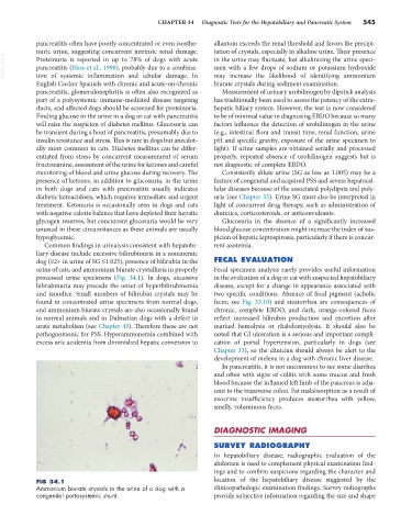
Common findings in urinalysis consistent with hepatobi- rent azotemia.
liary disease include excessive bilirubinuria in a nonanemic
dog (≥2+ in urine of SG ≤1.025), presence of bilirubin in the FECAL EVALUATION
urine of cats, and ammonium biurate crystalluria in properly Fecal specimen analysis rarely provides useful information
processed urine specimens (Fig. 34.1). In dogs, excessive in the evaluation of a dog or cat with suspected hepatobiliary
bilirubinuria may precede the onset of hyperbilirubinemia disease, except for a change in appearance associated with
and jaundice. Small numbers of bilirubin crystals may be two specific conditions. Absence of fecal pigment (acholic
found in concentrated urine specimens from normal dogs, feces; see Fig. 33.10) and steatorrhea are consequences of
and ammonium biurate crystals are also occasionally found chronic, complete EBDO, and dark, orange-colored feces
in normal animals and in Dalmatian dogs with a defect in reflect increased bilirubin production and excretion after
urate metabolism (see Chapter 43). Therefore these are not marked hemolysis or rhabdomyolysis. It should also be
pathognomonic for PSS. Hyperammonemia combined with noted that GI ulceration is a serious and important compli-
excess uric acidemia from diminished hepatic conversion to cation of portal hypertension, particularly in dogs (see
Chapter 33), so the clinician should always be alert to the
development of melena in a dog with chronic liver disease.
In pancreatitis, it is not uncommon to see some diarrhea
and often with signs of colitis with some mucus and fresh
blood because the inflamed left limb of the pancreas is adja-
cent to the transverse colon. Fat malabsorption as a result of
exocrine insufficiency produces steatorrhea with yellow,
smelly, voluminous feces.
DIAGNOSTIC IMAGING
SURVEY RADIOGRAPHY
In hepatobiliary disease, radiographic evaluation of the
abdomen is used to complement physical examination find-
ings and to confirm suspicions regarding the character and
FIG 34.1 location of the hepatobiliary disease suggested by the
Ammonium biurate crystals in the urine of a dog with a clinicopathologic examination findings. Survey radiographs
congenital portosystemic shunt. provide subjective information regarding the size and shape