
Page 572 - Small Animal Internal Medicine, 6th Edition
P. 572
544 PART IV Hepatobiliary and Exocrine Pancreatic Disorders
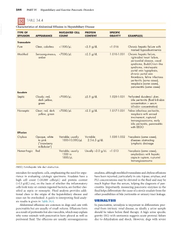
TABLE 34.4
VetBooks.ir Characteristics of Abdominal Effusion in Hepatobiliary Disease SPECIFIC
TYPE OF
PROTEIN
NUCLEATED CELL
EFFUSION APPEARANCE COUNT CONTENT GRAVITY EXAMPLE(S)
Transudate
Pure Clear, colorless <1500/µL <2.5 g/dL <1.016 Chronic hepatic failure with
marked hypoalbuminemia
Modified Serosanguineous, <7000/µL ≥2.5 g/dL 1.010-1.031 Chronic hepatic failure,
amber right-sided heart failure,
pericardial disease, caval
syndrome, Budd-Chiari–like
syndrome, intrahepatic
portal vein hypoplasia,
chronic portal vein
thrombosis, feline infectious
peritonitis (some cases),
neoplasia (some cases),
pancreatitis (some cases)
Exudate
Septic Cloudy; red, >7000/µL ≥2.5 g/dL 1.020-1.031 Perforated duodenal ulcer,
dark yellow, bile peritonitis (fluid bilirubin
green concentration > serum
bilirubin concentration)
Nonseptic Clear; red, dark >7000/µL ≥2.5 g/dL 1.017-1.031 Feline infectious peritonitis,
yellow, green neoplasia with serosal
involvement, ruptured
hemangiosarcoma, early
bile peritonitis, pancreatitis
with EBDO
Effusion
Chylous Opaque, white Variable; usually Variable; 1.030-1.032 Neoplasia (some cases),
to pink 1000-10,000/µL 2.5-6.5 g/dL diseases obstructing
(“strawberry lymphatic drainage
milkshake”)
Hemorrhagic Red Variable; usually Usually >3.0 g/dL <1.013 Neoplasia (some cases),
1500 to amyloidosis with hepatic
1000/µL capsule rupture, ruptured
hemangiosarcoma
EBDO, Extrahepatic bile duct obstruction.
mistaken for neoplastic cells, emphasizing the need for expe- exudates, although modified transudates and chylous effusions
rience in evaluating cytologic specimens. Exudates have a have been reported, particularly in cats. Lipase, amylase, and
high cell count (>20,000 cells/µL) and protein content PLI concentrations may be elevated in the fluid and may be
(>2.5 g/dL) and, on the basis of whether the inflammatory much higher than the serum, helping the diagnosis of pan-
cells look toxic or contain ingested bacteria, are further clas- creatitis. Importantly, measuring pancreatic enzymes in the
sified as septic or nonseptic. Fluid analysis provides addi- fluid helps differentiate the cause of a sterile exudate from the
tional clues to the origin of the hepatobiliary disease and other possibilities of bile peritonitis or urinary tract leakage.
must not be overlooked. A guide to interpreting fluid analy-
sis results is given in Table 34.4. URINALYSIS
Abdominal effusions are common in dogs and cats with In pancreatitis, urinalysis is important to differentiate prer-
pancreatitis but are usually of small volume. Effusions form enal from intrinsic renal disease, so ideally a urine sample
as a result of peritonitis but also vasculitis, which may explain should be taken before fluid therapy. A high urine specific
why some animals with pancreatitis have pleural as well as gravity (SG) with azotaemia suggests acute prerenal failure
peritoneal fluid. The effusions are usually serosanguineous due to dehydration and shock. However, dogs with severe