
Page 570 - Small Animal Internal Medicine, 6th Edition
P. 570
542 PART IV Hepatobiliary and Exocrine Pancreatic Disorders
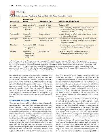
TABLE 34.3
VetBooks.ir Typical Clinicopathologic Findings in Dogs and Cats With Acute Pancreatitis—cont’d
CHANGES IN
PARAMETER DOGS CHANGES IN CATS CAUSE AND SIGNIFICANCE
Bilirubin Increased in 53% Increased in 64% Same as GGT
Cholesterol Increased in Increased in 64% Can be caused by cholestasis; unclear in others if
48%-80% cause or effect; often caused by concurrent or
predisposing disease
Triglycerides Commonly Rarely measured Unclear if cause or effect; often caused by concurrent
increased or predisposing disease
Neutrophils Increased in Increased in about 30%, Increase caused by inflammatory response; decrease
55%-60% decreased in 15% in some cats caused by consumption; may be poor
prognostic indicator
Hematocrit Increased in ≈20% As dogs Increase caused by dehydration; decrease caused by
and decreased anemia of chronic disease; gastrointestinal
in ≈20% ulceration
Platelets Commonly Usually normal Decrease caused by circulating proteases ±
decreased in disseminated intravascular coagulation
severe cases
(59%)
ALP, Alkaline phosphatase; ALT, alanine aminotransferase; AST, aspartate aminotransferase; GGT, γ-glutamyltranspeptidase.
Data from Schaer M: A clinicopathological survey of acute pancreatitis in 30 dogs and 5 cats, J Am Anim Hosp Assoc 15:681, 1979; Hill
RC et al: Acute necrotizing pancreatitis and acute suppurative pancreatitis in the cat: a retrospective study of 40 cases (1976-1989), J Vet
Intern Med 7:25, 1993; Hess RS et al: Clinicopathological, radiographic and ultrasonographic abnormalities in dogs with fatal acute
pancreatitis: 70 cases (1986-1995), J Am Vet Med Assoc 213:665, 1998; Mansfield CS et al: Review of feline pancreatitis. Part 2: clinical
signs, diagnosis and treatment, J Feline Med and Surgery 3:125, 2001.
combination of excessive renal and GI losses, reduced intake, size of red blood cells is reversible upon restoration of portal
and secondary hyperaldosteronism in dogs and cats with blood flow. If anemia is also present, microcytosis must be
severe chronic hepatobiliary disease. Metabolic alkalosis, distinguished from anemia of inflammatory disease, which
presumptive evidence of which might be an abnormally high can occasionally cause small red blood cells and relative iron
serum total carbon dioxide content confirmed by blood gas deficiency, or from iron deficiency anemia associated with
analysis, is usually caused by overzealous diuretic therapy in chronic GI blood loss, which might particularly be seen in a
dogs with chronic hepatic failure and ascites. Hypokalemia dog with chronic hepatitis and portal hypertension (see
and metabolic alkalosis potentiate each other and may also Chapter 36). The anemia sometimes seen in association with
worsen signs of hepatic encephalopathy (HE) by promoting the microcytosis of PSS is usually mild. A marked microcytic
the persistence of readily membrane-diffusible ammonia anemia greatly increases the index of suspicion for chronic
(NH 3 ). GI blood loss.
Strongly regenerative anemia, with macrocytosis, high
COMPLETE BLOOD COUNT reticulocyte count, and normal to slightly increased serum
There are few changes in blood cells that suggest hepatobili- protein concentration in a jaundiced dog, especially if
ary disease. Most are changes in erythrocytes associated with spherocytes are also identified, indicates hemolytic anemia
fragmentation or changes in cell size or membrane composi- and increased bilirubin formation as the cause of jaundice.
tion. Microcytosis (mean corpuscular volume [MCV] < 60 fL Cats and dogs with hemolytic anemia typically also have
in canine breeds other than the Japanese Akita or Shiba Inu), high serum liver enzyme and bile acid concentrations,
with normochromasia or slight hypochromasia (mean cell which indicate hepatic consequences developing secondary
hemoglobin concentration, 32-34 g/dL), is a common to the effects of marked hemolysis, such as hypoxia and
finding in dogs with congenital PSS (≥60%); it is less common thromboembolism.
in cats with congenital PSS (≤30%). Most affected animals Certain red blood cell morphologic changes are consis-
are not anemic. The cause of microcytosis, which has also tent with serious hepatobiliary disease and are related to
been observed with less frequency in dogs with chronic alterations in lipoprotein metabolism and irregularities in
hepatic failure and acquired PSS, is chelation of iron in the red blood cell membrane structure. Poikilocytes (abnormally
liver rather than absolute iron deficiency; therefore iron shaped red blood cells), such as acanthocytes (spiked red
supplementation does not help. However, the change in the blood cells), leptocytes (elongated, pale staining red blood