
Page 1405 - Veterinary Immunology, 10th Edition
P. 1405
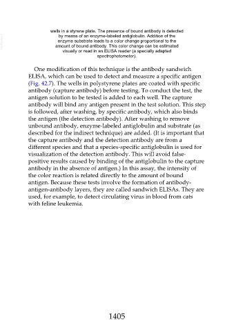
wells in a styrene plate. The presence of bound antibody is detected
  VetBooks.ir                amount of bound antibody. This color change can be estimated
                               by means of an enzyme-labeled antiglobulin. Addition of the
                              enzyme substrate leads to a color change proportional to the


                                 visually or read in an ELISA reader (a specially adapted
                                                  spectrophotometer).


                  One modification of this technique is the antibody sandwich

               ELISA, which can be used to detect and measure a specific antigen
               (Fig. 42.7). The wells in polystyrene plates are coated with specific
               antibody (capture antibody) before testing. To conduct the test, the
               antigen solution to be tested is added to each well. The capture
               antibody will bind any antigen present in the test solution. This step

               is followed, after washing, by specific antibody, which also binds
               the antigen (the detection antibody). After washing to remove
               unbound antibody, enzyme-labeled antiglobulin and substrate (as

               described for the indirect technique) are added. (It is important that
               the capture antibody and the detection antibody are from a
               different species and that a species-specific antiglobulin is used for
               visualization of the detection antibody. This will avoid false-
               positive results caused by binding of the antiglobulin to the capture

               antibody in the absence of antigen.) In this assay, the intensity of
               the color reaction is related directly to the amount of bound
               antigen. Because these tests involve the formation of antibody-

               antigen-antibody layers, they are called sandwich ELISAs. They are
               used, for example, to detect circulating virus in blood from cats
               with feline leukemia.


                                                        1405