
Page 311 - The Veterinary Laboratory and Field Manual 3rd Edition
P. 311
280 Susan C. Cork and Roy Halliwell
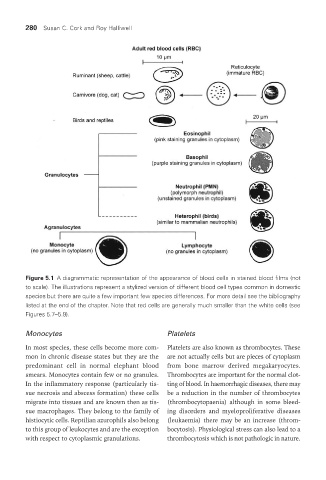
Figure 5.1 A diagrammatic representation of the appearance of blood cells in stained blood films (not
to scale). The illustrations represent a stylized version of different blood cell types common in domestic
species but there are quite a few important few species differences. For more detail see the bibliography
listed at the end of the chapter. Note that red cells are generally much smaller than the white cells (see
Figures 5.7–5.9).
Monocytes Platelets
In most species, these cells become more com- Platelets are also known as thrombocytes. These
mon in chronic disease states but they are the are not actually cells but are pieces of cytoplasm
predominant cell in normal elephant blood from bone marrow derived megakaryocytes.
smears. Monocytes contain few or no granules. Thrombocytes are important for the normal clot-
In the inflammatory response (particularly tis- ting of blood. In haemorrhagic diseases, there may
sue necrosis and abscess formation) these cells be a reduction in the number of thrombocytes
migrate into tissues and are known then as tis- (thrombocytopaenia) although in some bleed-
sue macrophages. They belong to the family of ing disorders and myeloproliferative diseases
histiocytic cells. Reptilian azurophils also belong (leukaemia) there may be an increase (throm-
to this group of leukocytes and are the exception bocytosis). Physiological stress can also lead to a
with respect to cytoplasmic granulations. thrombocytosis which is not pathologic in nature.
Vet Lab.indb 280 26/03/2019 10:25