
Page 313 - The Veterinary Laboratory and Field Manual 3rd Edition
P. 313
282 Susan C. Cork and Roy Halliwell
characteristics of the red and white blood cell Preparation and staining of blood and
population. It is recommended that a total bone marrow smears
white blood cell count is performed along with
a differential white cell count as this provides The preparation of a good blood film requires
the required context to help interpret the test the use of clean grease free slides (immersion
results. If it is difficult to collect a large volume of slides in 70% alcohol will remove grease but
of blood then it is acceptable to collect a small ensure that the slide is dry before use). A blood
amount in two capillary tubes for estimation of film can be made from a single drop of peripheral
PCV along with two blood smears to allow a blood or a drop from an EDTA sample submit-
rough assessment of the differential white cell ted in a vacutainer. The prepared film should
count and the red and white blood cell morphol- have a random distribution of white blood cells
ogy (see figures 5.6–5.9). throughout the film (Figure 5.2). Erythrocytes
Always ensure that the animal(s) to be should be distributed in a single layer on part
sampled are appropriately restrained. In some of the blood slide. A spreader is used for prep-
species it may be necessary to clip the hair in aration of smears, this should have a smooth,
the area around the vein to allow easy access. even edge with the corners cut to give a side
The vein may also be more easily visualized if slightly narrower than the width of the micro-
the skin is swabbed with alcohol or methylated scope slide. For examination of haemoparasites,
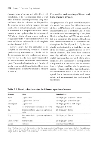
spirit. The usual collection site and the size of it is preferable to make thick and thin smears
needle recommended for collecting blood from from peripheral blood (see also the parasitology
common species of domestic animals is outlined section – Figure 3.36). Note that the haemato-
in Table 5.2. crit will affect the time it takes for the smear to
spread, that is, in anaemic animals it will spread
quickly and haemoconcentrated specimens will
take longer.
Table 5.2 Blood collection sites in different species of animal.
Species Site Needle size*
Horse Jugular vein 16–19 gauge/1.5–2 inch** length
Cow Jugular vein, tail vein 16–19 gauge/1.5–2 inch length
Sheep/goat Jugular vein 18–20 gauge/ 1.5–2 inch length
Pig Anterior thoracic vena cava, ear veins 20 gauge/1.5–4 inch
Cat Brachiocephalic, jugular or saphenous 20–25 gauge/1.5 inch
vein
Dog Brachiocephalic, jugular or saphenous 20–25 gauge/1.5 inch
vein
Small primate Femoral vein 20–26 gauge/1.5 inch
Mouse Tail vein Microhaematocrit tube
Note: *The larger the needle diameter, the smaller the gauge size, that is, a 16 gauge needle has a wider bore than a 25 gauge
needle. In general, you should try to use the largest gauge needle suitable as this reduces the risk of haemolysis when the
sample is drawn. **1 inch is equal to 25.4 mm.
Vet Lab.indb 282 26/03/2019 10:25