
Page 314 - The Veterinary Laboratory and Field Manual 3rd Edition
P. 314
Haematology 283
Method forward in a direct and even movement. The film
should be about 3–4 cm long. Air dry rapidly
Place a small drop of blood 1.5–2.0 cm from by waving the film in the air (this avoids crena-
one end of a clean slide. Hold the spreader at tion of the erythrocytes). Label the film either by
an angle of 30° in front of the drop of blood and writing with a pencil in the film itself or mark
bring it back to touch the blood (see Figures the slide using a diamond tipped pen. If staining
3.36 and 5.2a) allowing it to spread along the of the smear will be delayed by more than 24 h
edge. Complete the film by pushing the spreader
fix the film in methanol for 2 min, this should be
done as soon as the film is dry. If the films will
be stained within 24 h then air dry and place
(a)
out of the light and protect from dust. Air dry-
ing without fixation prior to staining is preferred
but not always possible in the field. If staining
(which includes a fixation step) will be delayed
by more than 24 h the film can deteriorate rap-
idly and so it is advisable to carry a small bottle
of methanol on field trips for this purpose. In the
laboratory, the slides can be stained with Giemsa
stain. This stain, along with Leishman stain
and Wright’s stain, is known as a Romanowsky
stain. There are a number of staining methods
(b)
recommended but the following has been found
satisfactory in our experience (see also Chapter
3). Several quick kit test stains are also available
for example, DiffQuick™.
Stain preparation
Prepare a stock Giemsa stain (Giemsa powder
1 g, glycerol 66 ml; mix thoroughly and heat to
56°C for 90 min, add 66 ml methanol. Mix thor-
oughly and leave to stand for 7 days; filter and
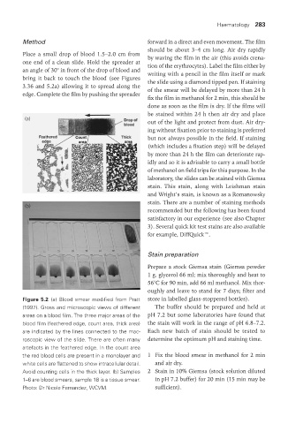
Figure 5.2 (a) Blood smear modified from Pratt store in labelled glass-stoppered bottles).
(1997). Gross and microscopic views of different The buffer should be prepared and held at
areas on a blood film. The three major areas of the pH 7.2 but some laboratories have found that
blood film (feathered edge, count area, thick area) the stain will work in the range of pH 6.8–7.2.
are indicated by the lines connected to the mac- Each new batch of stain should be tested to
roscopic view of the slide. There are often many determine the optimum pH and staining time.
artefacts in the feathered edge. In the count area
the red blood cells are present in a monolayer and 1 Fix the blood smear in methanol for 2 min
white cells are flattened to show intracellular detail. and air dry.
Avoid counting cells in the thick layer. (b) Samples 2 Stain in 10% Giemsa (stock solution diluted
1–6 are blood smears, sample 18 is a tissue smear. in pH 7.2 buffer) for 20 min (15 min may be
Photo: Dr Nicole Fernandez, WCVM. sufficient).
Vet Lab.indb 283 26/03/2019 10:25