
Page 398 - The Veterinary Laboratory and Field Manual 3rd Edition
P. 398
Pathology/cytology 367
and neurological). If neurological signs were anthrax spores which can contaminate an area
exhibited by the animal prior to death then a for a long period of time and potentially cause an
detailed examination of the brain and spinal outbreak of anthrax.
cord may be required but this is difficult and time During the post-mortem, it is helpful to have
consuming and not routinely done. If rabies is an assistant available to collect the samples and
suspected care must be taken to avoid human make a note of any gross abnormalities as the
exposure to the virus: wear gloves, a mask and, if examination proceeds (see example submission
available, use a designated kit to take the appro- form and full necropsy guidelines in Appendix
priate brain sections. Animal health staff likely 2). In some cases, the cause of death will be
to be exposed to rabies should be vaccinated to obvious from the clinical history or from previ-
protect them from rabies. If in any doubt about ous examinations of the animal, however, there
handling the case, take/send the whole head is often limited information available for a case.
(chilled) directly to the laboratory with a spe- In such circumstances, it is especially important
cial request to test for rabies. In cases of sudden to work from basic principles going through
death where anthrax is common take a periph- each body system in a systematic manner. In
eral blood smear (usually from the ear), stain it order to recognize an abnormality it is impor-
and examine for Gram +ve bacilli before open- tant to be familiar with the normal anatomy of
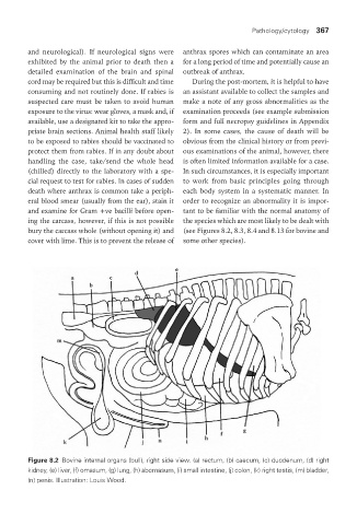
ing the carcass, however, if this is not possible the species which are most likely to be dealt with
bury the carcass whole (without opening it) and (see Figures 8.2, 8.3, 8.4 and 8.13 for bovine and
cover with lime. This is to prevent the release of some other species).
Figure 8.2 Bovine internal organs (bull), right side view. (a) rectum, (b) caecum, (c) duodenum, (d) right
kidney, (e) liver, (f) omasum, (g) lung, (h) abomasum, (i) small intestine, (j) colon, (k) right testis, (m) bladder,
(n) penis. Illustration: Louis Wood.
Vet Lab.indb 367 26/03/2019 10:26