
Page 399 - The Veterinary Laboratory and Field Manual 3rd Edition
P. 399
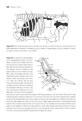
368  Susan C. Cork


            Figure 8.3  Bovine internal organs (cow), left side view. (a) rectum, (b) left kidney, (c) rumen, (d) costal line
            of the diaphragm, (e) reticulum, (f) heart, (g) lung, (h) spleen, (i) oesophagus, (j) ribs, (k) bladder, (m) ovary,
            (n) vagina, (o) uterus. Illustration: Louis Wood.


            Figure 8.4  A schematic representation
            of the topographical anatomy of a bird.
            There is quite a lot of anatomical varia-
            tion among avian species depending on
            their diet and evolutionary background.
            The diagram shown is based on the
            pigeon which is similar to domestic fowl.
            (No) nostril, (Tr) trachea, (Br) brain, (Sp)
            spinal cord (within vertebral column not
            shown),  (Oes)  oesophagus,  (H)  heart,
            (Lu) lungs, (Ki) kidney, (As) air sacs
            (these are located caudal and cranial to
            the lungs and act as ‘bellows’ to improve
            the respiratory efficiency of birds), note
            that most species of birds do not have a
            diaphragm. (Cr) crop (not all bird species
            have a crop, it acts to store food and in pigeons the lining secretes a ‘milk’ for chicks), (Pr) proventriculus
            and (Gi) gizzard or ventriculus (these form the stomach of the bird, this has a thick wall and often contains
            stones to help the bird break down food material as birds do not have teeth to grind fibrous food, the pro-
            ventriculus has a glandular lining), (Li) liver, (Ca) caecum (grain eating species have well developed paired
            caecae). (Int) small and large intestine. Some parasites such as coccidia can be partially identified by the
            region of the intestine or caecae that they prefer to invade. (Rec) rectum, (Cl) cloaca, birds do not have a
            separate faecal and urinary passage, both join to exit through the cloaca. Avian ‘faecal’ material contains
            the white urinary wastes (urates) and green/brown digestive wastes.


       Vet Lab.indb   368                                                                  26/03/2019   10:26