
Page 403 - The Veterinary Laboratory and Field Manual 3rd Edition
P. 403
372 Susan C. Cork
Respiratory system (see Figure 8.6) change. Note that in birds the lungs are fairly
small and fixed in place and there is no obvious
Examine the pleura which line the thoracic cav- diaphragm. Most of the breathing cycle in birds
ity before removing the trachea and lungs. Check involves the filling of the air sacs; examine the
that the diaphragm is still intact and look for main ones which lie in the abdominal cavity and
any signs of bruising on the inside of the rib the thoracic space. In healthy birds the air sacs
cage. Look for the presence of abnormal colou- should be clear like the pleural lining in mam-
ration, parasites and fluids in the airways. Note mals but in animals with respiratory diseases,
any areas of consolidation. Remove a piece of such as aspergillosis, the air sacs will be thick-
lung and see if it floats in water; if it sinks this ened and discoloured.
indicates consolidation rather than post-mortem
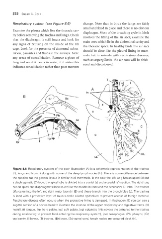
Figure 8.6 Respiratory system of the cow. Illustration (A) is a schematic representation of the trachea
(T), lungs and bronchi along with some of the deep lymph nodes (ln). There is some difference between
the species but the general layout is similar in all mammals. In the cow the left lung has an apical (a) and
a diaphragmatic (C) lobe, the apical lobe is divided into a cranial (a) and a caudal (a’) section. The right lung
has an apical and diaphragmatic lobe as well as the middle (b) lobe and the accessory (D) lobe. The trachea
bifurcates into the left and right major bronchi (B) and these branch into the bronchioles (b). The trachea
is lined with a protective layer of mucus and a ciliated epithelium to prevent access of foreign material.
Respiratory disease often occurs when the protective lining is damaged. In illustration (B) you can see a
sagittal section of a bovine head to illustrate the location of the upper respiratory and digestive tracts. (N)
nostril, (t) tongue, (hp) hard palate, (sp) soft palate, (ep) epiglottis (this covers the entrance to the trachea
during swallowing to prevent food entering the respiratory system), (oe) oesophagus, (Ph) pharynx, (Or)
oral cavity, (l) larynx, (T) trachea, (Br) brain, (Sc) spinal cord, lymph nodes are coloured black (ln).
Vet Lab.indb 372 26/03/2019 10:26